Quantitation of contacts among sensory, motor, and serotonergic neurons in the pedal ganglion of aplysia
- PMID: 14557611
- PMCID: PMC218004
- DOI: 10.1101/lm.63903
Quantitation of contacts among sensory, motor, and serotonergic neurons in the pedal ganglion of aplysia
Abstract
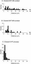
Present models of long-term sensitization in Aplysia californica indicate that the enhanced behavioral response is due, at least in part, to outgrowth of sensory neurons mediating defensive withdrawal reflexes. Presumably, this outgrowth strengthens pre-existing connections by formation of new synapses with follower neurons. However, the relationship between the number of sensorimotor contacts and the physiological strength of the connection has never been examined in intact ganglia. As a first step in addressing this issue, we used confocal microscopy to examine sites of contact between sensory and motor neurons in naive animals. Our results revealed relatively few contacts between physiologically connected cells. In addition, the number of contact sites was proportional to the amplitude of the EPSP elicited in the follower motor neuron by direct stimulation of the sensory neuron. This is the first time such a correlation has been observed in the central nervous system. Serotonin is the neurotransmitter most closely examined for its role in modulating synaptic strength at the sensorimotor synapse. However, the structural relationship of serotonergic processes and sensorimotor synapses has never been examined. Surprisingly, serotonergic processes usually made contact with sensory and motor neurons at sites located relatively distant from the sensorimotor synapse. This result implies that heterosynaptic regulation is due to nondirected release of serotonin into the neuropil.
Figures


Similar articles
-
Synaptic facilitation and behavioral dishabituation in Aplysia: dependence on release of Ca2+ from postsynaptic intracellular stores, postsynaptic exocytosis, and modulation of postsynaptic AMPA receptor efficacy.J Neurosci. 2005 Jun 8;25(23):5623-37. doi: 10.1523/JNEUROSCI.5305-04.2005. J Neurosci. 2005. PMID: 15944390 Free PMC article.
-
Serotonin release evoked by tail nerve stimulation in the CNS of aplysia: characterization and relationship to heterosynaptic plasticity.J Neurosci. 2002 Mar 15;22(6):2299-312. doi: 10.1523/JNEUROSCI.22-06-02299.2002. J Neurosci. 2002. PMID: 11896169 Free PMC article.
-
Differential effects of 4-aminopyridine, serotonin, and phorbol esters on facilitation of sensorimotor connections in Aplysia.J Neurophysiol. 1997 Jan;77(1):177-85. doi: 10.1152/jn.1997.77.1.177. J Neurophysiol. 1997. PMID: 9120559
-
Widespread anatomical projections of the serotonergic modulatory neuron, CB1, in Aplysia.Invert Neurosci. 1995;1(2):173-83. doi: 10.1007/BF02331914. Invert Neurosci. 1995. PMID: 9372141
-
Postsynaptic regulation of the development and long-term plasticity of Aplysia sensorimotor synapses in cell culture.J Neurobiol. 1994 Jun;25(6):666-93. doi: 10.1002/neu.480250608. J Neurobiol. 1994. PMID: 8071666 Review.
Cited by
-
Synaptic plasticity in myenteric neurons of the guinea-pig distal colon: presynaptic mechanisms of inflammation-induced synaptic facilitation.J Physiol. 2007 Jun 1;581(Pt 2):787-800. doi: 10.1113/jphysiol.2007.128082. Epub 2007 Mar 15. J Physiol. 2007. PMID: 17363386 Free PMC article.
-
Synaptic facilitation and behavioral dishabituation in Aplysia: dependence on release of Ca2+ from postsynaptic intracellular stores, postsynaptic exocytosis, and modulation of postsynaptic AMPA receptor efficacy.J Neurosci. 2005 Jun 8;25(23):5623-37. doi: 10.1523/JNEUROSCI.5305-04.2005. J Neurosci. 2005. PMID: 15944390 Free PMC article.
-
Regulation of behavioral and synaptic plasticity by serotonin release within local modulatory fields in the CNS of Aplysia.J Neurosci. 2006 Dec 6;26(49):12682-93. doi: 10.1523/JNEUROSCI.3309-06.2006. J Neurosci. 2006. PMID: 17151271 Free PMC article.
-
Cellular-resolution gene expression mapping reveals organization in the head ganglia of the gastropod, Berghia stephanieae.J Comp Neurol. 2024 Jun;532(6):e25628. doi: 10.1002/cne.25628. J Comp Neurol. 2024. PMID: 38852042 Free PMC article.
-
Long-lasting synaptic potentiation induced by depolarization under conditions that eliminate detectable Ca2+ signals.J Neurophysiol. 2010 Mar;103(3):1283-94. doi: 10.1152/jn.00704.2009. Epub 2009 Dec 30. J Neurophysiol. 2010. PMID: 20042699 Free PMC article.
References
-
- Bailey, C.H. and Chen, M. 1983. Morphological basis of long-term habituation and sensitization in Aplysia. Science 220: 91–93. - PubMed
-
- Bailey, C.H., Thompson, E.B., Castellucci, V.F., and Kandel, E.R. 1979. Ultrastructure of the synapses of sensory neurons that mediate the gill-withdrawal reflex in Aplysia. J. Neurocytol. 8: 415–444. - PubMed
Publication types
MeSH terms
Substances
Grants and funding
LinkOut - more resources
Full Text Sources
Research Materials
