. 2003 Nov;77(21):11312-23.
doi: 10.1128/jvi.77.21.11312-11323.2003.

Coronaviruses as vectors: position dependence of foreign gene expression

Affiliations

Coronaviruses as vectors: position dependence of foreign gene expression

Cornelis A M de Haan et al. J Virol. 2003 Nov.

Abstract

Coronaviruses are the enveloped, positive-stranded RNA viruses with the largest RNA genomes known. Several features make these viruses attractive as vaccine and therapeutic vectors: (i) deletion of their nonessential genes is strongly attenuating; (ii) the genetic space thus created allows insertion of foreign information; and (iii) their tropism can be modified by manipulation of the viral spike. We studied here their ability to serve as expression vectors by inserting two different foreign genes and evaluating systematically the genomic position dependence of their expression, using a murine coronavirus as a model. Renilla and firefly luciferase expression cassettes, each provided with viral transcription regulatory sequences (TRSs), were inserted at several genomic positions, both independently in different viruses and combined within one viral genome. Recombinant viruses were generated by using a convenient method based on targeted recombination and host cell switching. In all cases high expression levels of the foreign genes were observed without severe effects on viral replication in vitro. The expression of the inserted gene appeared to be dependent on its genomic position, as well as on the identity of the gene. Expression levels increased when the luciferase gene was inserted closer to the 3' end of the genome. The foreign gene insertions generally reduced the expression of upstream viral genes. The results are consistent with coronavirus transcription models in which the transcription from upstream TRSs is attenuated by downstream TRSs. Altogether, our observations clearly demonstrate the potential of coronaviruses as (multivalent) expression vectors.


Figures

FIG. 1.
FIG. 1.
Plasmid constructs. The transcription vectors from which the defective RNAs were produced in vitro by using T7 RNA polymerase are shown. Vector pMH54 has been described before (24). The other vectors were derived from pMH54 as described in Materials and Methods. The arrow at the left end of each vector indicates the T7 promoter; the solid circle represents the polylinker between the 5′-end segment of the MHV genome (denoted 5′/1) and either the HE gene or the 3′ end of the replicase gene (1b), followed by the structural and group-specific genes and the 3′-untranslated region and the polyadenylated segment (denoted together as 3′/A). The “m” indicates a 126-nt segment from the 3′ end of the M gene. Junctions between viral and insert sequences are marked by numbers in circles; the actual sequences are shown in Fig. 2. The recombinant viruses generated with the transcription vectors are indicated at the right.
FIG. 2.
FIG. 2.
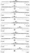
Sequences at the junctions of the inserted luciferase gene cassettes. Shown are the sequences of the junctions indicated by the circled numbers in Fig. 1. The sequence of the expression cassette is in boldface. Nucleotides outside the expression cassette that differ from the original wild-type MHV-A59 sequence are underlined and in boldface.
FIG. 3.
FIG. 3.
Recombinant viruses with luciferase gene cassettes between the E and the M gene. (A) Genomic organization of the recombinant wild-type MHV-A59 (MHV-WT) and of the recombinants containing either an RL or an FL expression cassette between the E and M genes. (B) Intracellular RNA synthesis by the recombinant MHVs was analyzed as described in Materials and Methods. Purified total cytoplasmic RNA was separated by electrophoresis through 1% agarose containing formaldehyde, and labeled RNA was visualized by fluorography. The position of each RNA species is indicated by triangles alongside the panels. An I, II, or III in parentheses indicates the viral source of the RNA species as being MHV-WT, MHV-ERLM, or MHV-EFLM, respectively. (C) Single-step growth kinetics of the MHV recombinants. LR7 cells were infected with each recombinant MHV at an MOI of 8. Viral infectivity in the culture media at different times postinfection was determined by a quantal assay on LR7 cells, and TCID50 values were calculated. Independently generated recombinants are indicated by the addition of A and B. (D) In a parallel experiment, the intracellular expression of RL and FL was determined at different times postinfection by using a luminometer (in RLU).
FIG. 4.
FIG. 4.
PCR and sequence analysis of aberrant sgRNAs. RT-PCR was used to amplify regions of cytoplasmic RNA isolated from cells infected with MHV-EFLM (A) or MHV-ERLM (B). The approximate locations of primers 1092 (9), 1814 (5′-GGTACTTCGTCCACAAACAC-3′), 1828 (5′-GGCGAAGAAGGAGAATAGG-3′), and 1855 (5′-GAGAACTCGCTCAACGAAC-3′) in the recombinant MHV genomes are shown. Primers 1092 and 1814 were used in the RT steps, whereas primer pairs 1814-1495, 1828-1495, and 1855-1495 were used for the PCR. The 3′ end of primer 1495 (5′-CCCGGGATCCATTTAGGTGACACTATAGAATATAAGAGTGATTGGCGTCC-3′) corresponds to the leader sequence of MHV. FLi and RLi indicate the intended leader-to-body fusion sites located just 5′ upstream of the FL and RL gene, respectively. FL*, FL**, and RL* indicate the aberrant leader-to-body fusion sites. The homology between the sequence of the fusion site (upper sequence) and the 5′ end of the genome (lower sequence) is marked by vertical bars. The arrows below the sequences indicate the sites of fusion of the body sequence to the leader sequence of the sgRNA.
FIG. 5.
FIG. 5.
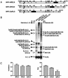
Recombinant viruses with RL gene cassettes at different positions. (A) The genomic organization of the recombinant viruses containing the RL gene cassette between the M and the N genes (MHV-MRLN), between the E and the M genes (MHV-ERLM), and between the 2a and S genes is shown. (B) Intracellular RNA synthesis by the recombinant MHVs was analyzed as described in Materials and Methods and in the legend to Fig. 3. The position of each RNA species is indicated by triangles alongside the panels. An I, II, or III in parentheses indicates the viral source of the RNA species, i.e., MHV-WT, MHV-ERLM, or MHV-EFLM, respectively. (C) Titers and luciferase expression levels of the MHV recombinants at 9 h postinfection. LR7 cells were infected in quadruplicate with each recombinant MHV at an MOI of 8. Viral infectivity in the culture media was determined by a quantal assay on LR7 cells, and TCID50 were calculated. In the same experiment the intracellular expression of RL (in RLU) was determined by using a luminometer; standard deviations are indicated.
FIG. 6.
FIG. 6.
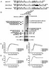
Viral protein synthesis by different luciferase-expressing MHV recombinants. LR7 cells were infected with recombinant viruses and subsequently labeled with 35S-labeled amino acids from 5 to 8 h postinfection. (A) Total lysates of cells and culture media were prepared and used for immunoprecipitation with the anti-MHV serum K134, and the precipitates were analyzed by SDS-15% PAGE. An example is shown for MHV-WT. The positions of the different viral proteins are indicated on the left, while those of molecular mass marker proteins are indicated on the right of the gel. (B) For quantitative analysis, the amounts of radioactivity in the M, S, and N proteins were determined by phosphorimager scanning of the dried gels from three independent experiments, with the indicated recombinant viruses. The ratios of the amounts of the S and N (S/N), M and N (M/N), and S and M (S/M) proteins synthesized in the cells infected with the luciferase-expressing viruses were calculated relative to those in MHV-WT-infected cells (set at 1.0); the standard deviations are indicated.
FIG. 7.
FIG. 7.
Expression of RL from a virus with a rearranged genome. (A) Genomic organization of two recombinant viruses each containing the RL gene between the E and M genes: one in the context of a “normal” genome (MHV-ERLM) and the other in a genomically rearranged background (MHV-ERLMSmN). The “m” indicates a 126-nt segment from the 3′ end of the M gene. (B and C) LR7 cells were infected with each recombinant MHV at an MOI of 8. Viral infectivity in the culture media was determined by a quantal assay on LR7 cells either at different times (B, left graph) or at 9 h (C, left graph) postinfection, the latter in quadruple, and TCID50 values were calculated. In the same experiment, the intracellular expression of RL was determined (in RLU) at the same times postinfection (B and C, middle graphs) by using a luminometer. The right panels of B and C show the RL expression as calculated relative to the virus titer (i.e., RLU/TCID50). The standard deviations are indicated.
FIG. 8.
FIG. 8.
Expression of RL and FL from a single genome. (A) The genomic organization of the recombinant viruses containing the RL gene between the 2a and S genes (MHV-2aRLS), the FL gene between the E and M genes (MHV-EFLM), or a combination thereof (MHV-RLFL) or containing the FL gene between the 2a and the S genes and the RL gene between the E and the M genes (MHV-FLRL) is shown. (B) Titers and luciferase expression of the MHV recombinants at 9 h postinfection. LR7 cells were infected in duplicate with each recombinant MHV at an MOI of 8. Viral infectivity in culture media was determined by a quantal assay on LR7 cells, and the TCID50 values were calculated. In the same experiment the intracellular expression of RL and FL was also determined (in RLU). (C) Ratio of RL and FL luciferase expression (RL/FL ratio) of MHV-RLFL and MHV-FLRL. LR7 cells were infected as described above. At 8 h postinfection, the intracellular expression of RL and FL was determined, and the RL/FL ratio was calculated. The standard deviations are indicated.

Similar articles

Cited by

References

    1. Almazan, F., J. M. Gonzalez, Z. Penzes, A. Izeta, E. Calvo, J. Plana-Duran, and L. Enjuanes. 2000. Engineering the largest RNA virus genome as an infectious bacterial artificial chromosome. Proc. Natl. Acad. Sci. USA 97:5516-5521. - PMC - PubMed
    1. Alonso, S., A. Izeta, I. Sola, and L. Enjuanes. 2002. Transcription regulatory sequences and mRNA expression levels in the coronavirus transmissible gastroenteritis virus. J. Virol. 76:1293-1308. - PMC - PubMed
    1. An, S., and S. Makino. 1998. Characterizations of coronavirus cis-acting RNA elements and the transcription step affecting its transcription efficiency. Virology 243:198-207. - PMC - PubMed
    1. Bredenbeek, P. J., C. J. Pachuk, A. F. Noten, J. Charite, W. Luytjes, S. R. Weiss, and W. J. Spaan. 1990. The primary structure and expression of the second open reading frame of the polymerase gene of the coronavirus MHV-A59; a highly conserved polymerase is expressed by an efficient ribosomal frameshifting mechanism. Nucleic Acids Res. 18:1825-1832. - PMC - PubMed
    1. Casais, R., V. Thiel, S. G. Siddell, D. Cavanagh, and P. Britton. 2001. Reverse genetics system for the avian coronavirus infectious bronchitis virus. J. Virol. 75:12359-12369. - PMC - PubMed

Publication types

MeSH terms

LinkOut - more resources