. 2003 Nov;77(21):11332-46.
doi: 10.1128/jvi.77.21.11332-11346.2003.

Measles virus (MV) nucleoprotein binds to a novel cell surface receptor distinct from FcgammaRII via its C-terminal domain: role in MV-induced immunosuppression

Affiliations

Measles virus (MV) nucleoprotein binds to a novel cell surface receptor distinct from FcgammaRII via its C-terminal domain: role in MV-induced immunosuppression

David Laine et al. J Virol. 2003 Nov.

Abstract

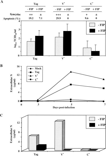
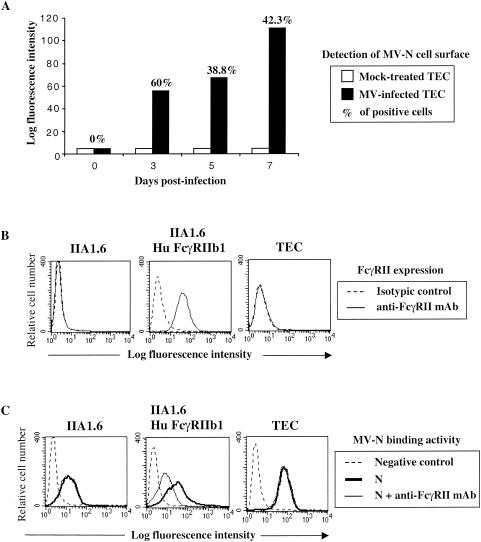
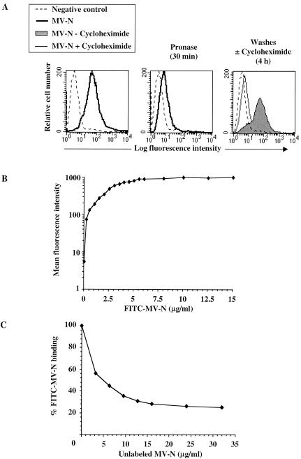
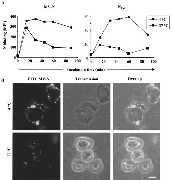
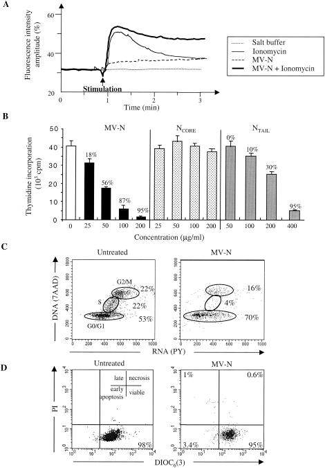
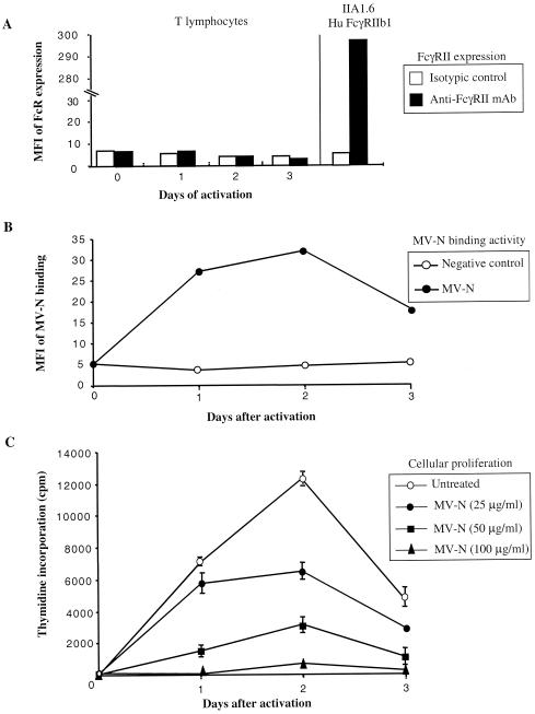
During acute measles virus (MV) infection, an efficient immune response occurs, followed by a transient but profound immunosuppression. MV nucleoprotein (MV-N) has been reported to induce both cellular and humoral immune responses and paradoxically to account for immunosuppression. Thus far, this latter activity has been attributed to MV-N binding to human and murine FcgammaRII. Here, we show that apoptosis of MV-infected human thymic epithelial cells (TEC) allows the release of MV-N in the extracellular compartment. This extracellular N is then able to bind either to MV-infected or uninfected TEC. We show that recombinant MV-N specifically binds to a membrane protein receptor, different from FcgammaRII, highly expressed on the cell surface of TEC. This new receptor is referred to as nucleoprotein receptor (NR). In addition, different Ns from other MV-related morbilliviruses can also bind to FcgammaRII and/or NR. We show that the region of MV-N responsible for binding to NR maps to the C-terminal fragment (N(TAIL)). Binding of MV-N to NR on TEC triggers sustained calcium influx and inhibits spontaneous cell proliferation by arresting cells in the G(0) and G(1) phases of the cell cycle. Finally, MV-N binds to both constitutively expressed NR on a large spectrum of cells from different species and to human activated T cells, leading to suppression of their proliferation. These results provide evidence that MV-N, after release in the extracellular compartment, binds to NR and thereby plays a role in MV-induced immunosuppression.


Figures

FIG. 1.
FIG. 1.
Extracellular MV-N release after apoptosis of MV-infected cells. TEC were infected with recombinant MV Tag/Edm, MV-V, or MV-C and then treated with or without the FIP Z-d-Phe-l-Phe-Gly-OH. (A) Inhibition of MV-induced cytopathic effect in TEC by FIP on day 5. Attached TEC were visualized by May-Grunwald-Giemsa staining, and syncytium formation was determined under light microscope. ±, +, ++, and +++, relative intensities of the cytopathic effect as defined by the number and the size of the syncytia. The percentage of pooled attached and floating TEC was evaluated by measuring caspase activity by flow cytometry. The number of infectious virus particles produced (TCID50/milliliter) is expressed as the mean ± the standard deviation (SD) of three different experiments. (B) Time course determination of extracellular N release in MV-infected TEC culture supernatants. Samples were prepared and assayed by ELISA as described in Materials and Methods. N concentrations were evaluated with known amounts of standard purified viral MV-N. (C) Inhibition of extracellular N release by FIP on day 5. The experiments were performed twice, and SD values were <10%.
FIG. 2.
FIG. 2.
Binding of MV-N to cells expressing FcγRII and another cell surface receptor. (A) Cell surface expression of N on living MV-infected TEC. TEC were infected with Edmonston MV strain at 0.1 PFU/cell. At different times postinfection, MV-N cell surface detection was determined by flow cytometry analysis with anti-MV-N (Cl25) and streptavidin-PE. The results are representative of six different experiments. (B) FcγRII expression on murine IIA1.6, on IIA1.6 expressing human FcγRIIb1, and on human TEC cell lines was detected by using anti-CD32-PE. For IIA1.6 and TEC, isotypic control and anti-CD32-PE are totally superimposed. (C) MV-N binding was detected with specific biotinylated anti-MV-N (Cl25) and then revealed with streptavidin-PE prior to flow cytometry analysis. Cells were incubated with 5 μg of purified recombinant MV-N in the absence (heavy line) or in the presence (thin line) of human blocking KB61 MAb. As a negative control, cells were incubated without MV-N in the presence of anti-MV-N (Cl25) and streptavidin-PE (dotted line). For IIA1.6 and TEC, the fluorescence intensity obtained in the presence of KB61 MAb superimposes on that obtained in the absence of this MAb. The results are representative from one of three independent experiments.
FIG. 3.
FIG. 3.
Specific binding of MV-N to a protein receptor distinct from FcγRII. (A) Requirement of protein synthesis for recovery of MV-N-binding activity after protease treatment. TEC were untreated (left panel) or treated with pronase (middle panel) before MV-N binding. Pronase-treated TEC were incubated for 4 h at 37°C in the absence (gray histogram) or in the presence of cycloheximide (thin line), prior to MV-N binding (right panel). As a negative control, cells were incubated with specific anti-MV-N (Cl25) MAb and streptavidin-PE in the absence of MV-N (dotted line). (B) Dose-dependent binding of FITC-labeled MV-N. TEC were incubated 1 h at 4°C with various amounts of FITC-MV-N, followed by extensive washes prior to flow cytometry analysis. (C) Competition between unlabeled MV-N and FITC-MV-N. Cells were incubated with FITC-MV-N (2.5 μg/well, corresponding to 50% of binding) and increasing amounts of unlabeled MV-N. Percentages of labeled MV-N binding were reported and calculated assuming that the MFI observed with FITC-MV-N alone is 100%. The results are representative from one of three independent experiments.
FIG. 4.
FIG. 4.
Morbillivirus-N and NTAIL binding to cell surface receptor(s). (A) Binding of Morbillivirus-N to different cell lines expressing or not expressing FcγRII. The extent of binding has been evaluated as described in Materials and Methods. The MFI is indicated as follows: −, no binding; +, ++, and +++, MFI values of ca. 10, between 10 and 100, and >100, respectively. The results are representative of three to six independent experiments. (B) At the top of the panel is shown MV-N domain purification from bacteria. Purified proteins were separated by sodium dodecyl sulfate-15% polyacrylamide gel electrophoresis and stained with Coomassie brillant blue. M, molecular mass markers. At the bottom of the panel is shown MV-N domain organization. MV-N (aa 1 to 525) is divided into two domains: NCORE (aa 1 to 400) and NTAIL (aa 401 to 525). The epitopes recognized by the anti-MV-N Cl120 and Cl25 MAbs are indicated. N-N and N-RNA binding sites are also shown. (C) Binding of MV NTAIL, but not NCORE, to NR. TEC were incubated with 5 μg of purified MV-tagged N (thin line) prior to flow cytometry analysis. Binding of NCORE and NTAIL (5 μg, heavy line) was detected by using biotinylated anti-MV-N Cl120 (left panel) and Cl25 (right panel), respectively, and then revealed with streptavidin-PE. As a negative control, neither N nor N domains were added, and the cell lines were incubated in the presence of specific biotinylated anti-N MAb and streptavidin-PE (dotted line). The results are representative from one of three independent experiments. (D) Specific NTAIL binding to NR. Competition binding experiments were performed by incubating MV-N (2.5 μg, corresponding to 50% of binding) with increasing amounts of either NTAIL or NCORE. Bacterially purified MV-N binding was revealed by biotinylated anti-MV-N Cl120 or Cl25 MAbs, depending upon whether NTAIL or NCORE was used as a competitor, respectively. The percentage of MV-N binding inhibition was calculated assuming that the MFI observed with MV-N alone represents 0% of inhibition. The results are representative from one to three independent experiments.
FIG. 5.
FIG. 5.
MV-N constitutive internalization in TEC. (A) Downregulation of MV-N binding in TEC. MV-N binding to TEC was measured at either 4 or 37°C by flow cytometry. Binding of recombinant MV-N (left panel) and NTAIL (right panel) was revealed by using anti-MV-N (Cl25) and streptavidin-PE. The results are representative of three independent experiments (SD values were <15%). (B) MV-N internalization at 37°C in vesicle-like structures within the cytoplasm. FITC-MV-N was used for confocal microscopy analysis. Single confocal sections show FITC fluorescence (left panel), transmission (middle panel), and overlap of both (right panel). Pictures are taken on TEC at either 4°C (upper panel) or 37°C (lower panel) after 1 h of incubation. Shown are representative results from one of three independent experiments. The white scale bar shown in the lower right panel corresponds to 5 μm.
FIG. 6.
FIG. 6.
NR is functional upon MV-N binding. (A) MV-N sustains ionomycin-mediated Ca2+ mobilization in TEC. At the time indicated by the arrow, 2 ml of loaded TEC (2 × 10⁵/ml) was stimulated with salt buffer, MV-N (40 μg/ml), and/or ionomycin (10−5 M). Cytosolic Ca2+ influx was then measured. (B) Effect of MV-N or NTAIL on spontaneous TEC proliferation. TEC were incubated with medium alone (open bar, control) and with various amounts of bacterially purified recombinant MV-N (solid bars), NCORE (dotted bars), or NTAIL (gray bars). Each value represents mean ± the SD of triplicate cultures. (C) Cell cycle analysis of TEC with 7AAD(DNA)/PY(RNA) staining. Two-dimensional analysis of DNA/RNA content is shown. The percentages of cells in the G0/G1, S, and G2/M phases of the cell cycle are shown. TEC were untreated or treated for 2 days with 200 μg of MV-N/ml purified from insect cells. (D) Viability and apoptosis were analyzed by flow cytometry in untreated TEC and in MV-N-treated TEC as in panel C after DIOC6/PI double staining. Viable cells are DIOC6 positive and PI negative. In contrast, apoptotic cells have both a decrease of mitochondrial transmembrane potential and permeabilized membranes that render them DIOC6 negative and PI positive, respectively. The results are from one representative experiment out of three.
FIG. 7.
FIG. 7.
Inhibition of human activated T-cell proliferation after MV-N binding to NR. (A) FcγRII expression on human activated T cells was detected by using anti-CD32-PE at different times after stimulation. (B) MV-N binding was detected with specific biotinylated anti-MV-N (Cl25) and then revealed with streptavidin-PE prior to flow cytometry analysis. Cells were incubated with 5 μg of purified recombinant MV-N from insect cells (•). As a negative control, cells were incubated without MV-N in the presence of anti-MV-N (Cl25) and streptavidin-PE (○). The experiments were performed twice, and the SD values were <10%. (C) Effect of MV-N on human activated T-cell proliferation. Activated T cells were incubated with either medium alone (○) or with various amounts of recombinant MV-N from insect cells (•, ▪, and ▴) for 24, 48, or 72 h. Each value represents the mean ± SD of triplicate cultures. The results are from one representative experiment out of three.

Similar articles

Cited by

References

    1. Auwaerter, P. G., H. Kaneshima, J. M. McCune, G. Wiegand, and D. E. Griffin. 1996. Measles virus infection of thymic epithelium in the SCID-hu mouse leads to thymocyte apoptosis. J. Virol. 70:3734-3740. - PMC - PubMed
    1. Bankamp, B., S. M. Horikami, P. D. Thompson, M. Huber, M. Billeter, and S. A. Moyer. 1996. Domains of the measles virus N protein required for binding to P protein and self-assembly. Virology 216:272-277. - PubMed
    1. Barrett, T. 1999. Morbillivirus infections, with special emphasis on morbilliviruses of carnivores. Vet. Microbiol. 69:3-13. - PubMed
    1. Bhella, D., A. Ralph, L. B. Murphy, and R. P. Yeo. 2002. Significant differences in nucleocapsid morphology within the Paramyxoviridae. J. Gen. Virol. 83:1831-1839. - PubMed
    1. Bikle, D. D., A. Ratnam, T. Mauro, J. Harris, and S. Pillai. 1996. Changes in calcium responsiveness and handling during keratinocyte differentiation: potential role of the calcium receptor. J. Clin. Investig. 97:1085-1093. - PMC - PubMed

Publication types

MeSH terms

LinkOut - more resources