. 2003 Nov;77(21):11633-43.
doi: 10.1128/jvi.77.21.11633-11643.2003.

Murine cytomegalovirus m41 open reading frame encodes a Golgi-localized antiapoptotic protein

Affiliations

Murine cytomegalovirus m41 open reading frame encodes a Golgi-localized antiapoptotic protein

Wolfram Brune et al. J Virol. 2003 Nov.

Abstract

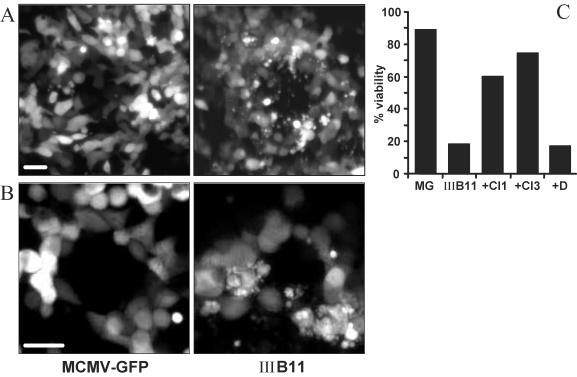
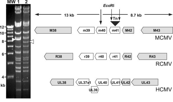
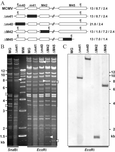
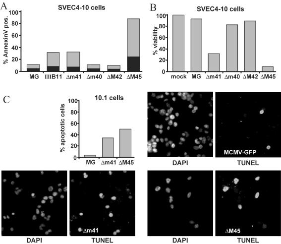
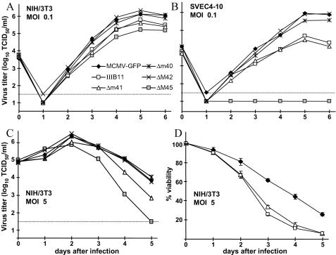
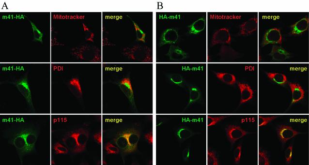
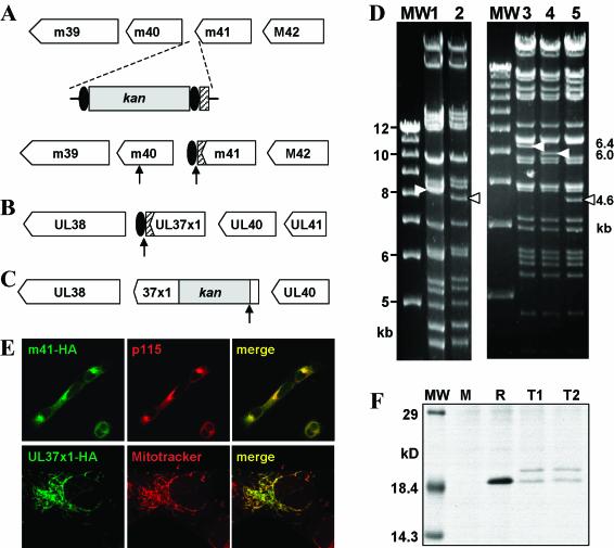
Viruses have evolved various strategies to prevent premature apoptosis of infected host cells. Some of the viral genes mediating antiapoptotic functions have been identified by their homology to cellular genes, but others are structurally unrelated to genes of known function. In this study, we used a random, unbiased approach to identify such genes in the murine cytomegalovirus genome. From a library of random transposon insertion mutants, a mutant virus that caused premature cell death was isolated. The transposon was inserted within open reading frame m41. An independently constructed m41 deletion mutant showed the same phenotype, whereas deletion mutants lacking the adjacent genes m40 and M42 did not. Apoptosis occurred in different cell types, could be blocked by caspase inhibitors, and did not require p53. Within the murine cytomegalovirus genome, m41, m40, and m39 form a small cluster of genes of unknown function. They are homologous to r41, r40, and r39 of rat cytomegalovirus, but lack sequence homology to UL41, UL40, and UL37 exon 1 (UL37x1) which are located at the corresponding positions of the human cytomegalovirus genome. Unlike UL37x1 of human cytomegalovirus, which encodes a mitochondrion-localized inhibitor of apoptosis that is essential for virus replication, m41 encodes a protein that localizes to the Golgi apparatus. The murine cytomegalovirus m41 product is the first example of a Golgi-localized protein that prevents premature apoptosis and thus extends the life span of infected cells.


Figures

FIG. 1.
FIG. 1.
Apoptotic cell death induced by an MCMV transposon insertion mutant. (A) NIH 3T3 and (B) SVEC4-10 cells were infected at a multiplicity of infection of 0.01 TCID50/cell with mutant IIIB11 and with the control virus, MCMV-GFP. The fluorescent images show virus plaques with morphological signs of apoptosis (membrane blebbing, disintegration of cells with formation of apoptotic bodies) in individual IIIB11-infected cells, most prominently seen around the center of the plaque. Bar, 20 μm. (C) The viability of NIH 3T3 cells infected with IIIB11 was markedly reduced compared to that of cells infected with MCMV-GFP (MG) 36 h after infection at a multiplicity of 10 TCID50/cell. Cell death could be largely inhibited by treating cells with caspase inhibitor I or III (CI1 or CI3, i.e., Z-VAD-FMK or Boc-D-FMK, respectively) but not by addition of dimethyl sulfoxide (D), the solvent used for the caspase inhibitors.
FIG. 2.
FIG. 2.
Location of the transposon insertion in mutant IIIB11. The transposon insertion within ORF m41 of the MCMV genome results in a change of the EcoRI restriction pattern (white arrowheads) in IIIB11 BAC (lane 2) compared to the MCMV wild-type BAC (lane 1), because the transposon (Tn) contains EcoRI sites within its inverted repeats. The exact location of the transposon insertion was determined by sequencing. ORFs m39, m40, and m41 show no sequence homology to the HCMV ORFs at the corresponding positions but are homologous to r39 to r41, respectively, of rat CMV (RCMV). Genes conserved among the three CMVs are shown in gray. Lane MW, molecular size markers.
FIG. 3.
FIG. 3.
Construction of MCMV deletion mutants. (A) ORFs m41, m40, M42, and M45 were deleted from the MCMV-GFP BAC (MG) and replaced with a zeo gene (black box). This resulted in loss or gain of an EcoRI restriction site (E) in mutants MCMVΔm40, ΔM42, and ΔM45 but not in Δm41. The calculated sizes of the EcoRI restriction fragments in the m40 to M45 region are given on the right. (B) Restriction patterns visualized by ethidium bromide staining. Bands with sizes different from that of the parental MCMV-GFP BAC are indicated by arrowheads. An SnaBI restriction site within m41 is lost in MCMVΔm41, resulting in fusion of two SnaBI fragments of 4.8 and 3.3 kb into a new fragment of 8.1 kb (arrowheads). (C) Southern blot of EcoRI-digested BACs, hybridized with a probe specific for the zeo gene sequence. Lane MW, molecular size markers.
FIG. 4.
FIG. 4.
MCMV deletion mutations cause apoptosis. SVEC4-10 endothelial cells were infected at a multiplicity of 5 TCID50/cell with MCMV-GFP (MG) or deletion mutants. (A) Phosphatidylserine on the outer leaflet of the cell membrane was detected with annexin V at 24 h after infection. The percentage of cells staining positive with both annexin V and propidium iodide (indicating late-stage apoptosis or nonapoptotic cell death) is shown in black. (B) Cell viability was determined 32 h after infection by the neutral red inclusion assay. The viability of cells infected with the Δm41 or the ΔM45 mutant was markedly reduced, whereas cells infected with MCMVΔm40 or MCMVΔM42 were as viable as cells infected with MCMV-GFP. (C) Cells of p53-deficient fibroblast cell line 10.1 also underwent apoptosis after infection with the Δm41 or the ΔM45 mutant. Cells were stained on coverslips by the TUNEL assay and counterstained with DAPI. Fewer cells are seen in the photomicrographs of cells infected with the Δm41 or ΔM45 mutant virus because dead and disintegrated cells detached from the coverslips.
FIG. 5.
FIG. 5.
Growth properties of MCMV mutants. (A) NIH 3T3 fibroblasts and (B) SVEC4-10 endothelial cells were infected at a multiplicity of 0.1 TCID50/cell for multistep growth curves. Deletion and insertion mutagenesis of the m41 ORF (mutants MCMVΔm41 and IIIB11) had no obvious effect on virus growth in NIH 3T3 fibroblasts and resulted in only moderately reduced titers in SVEC4-10 endothelial cells. By contrast, the ΔM45 mutant failed to grow on SVEC4-10 cells, consistent with previous results (6). (C) After infection at a multiplicity of 5 TCID50/cell, the titers of the Δm41 and the ΔM45 mutants declined faster than the titers of the control viruses. All titers represent mean values of two or three parallel experiments. Dotted line, detection limit. (D) A time course analysis showed a faster decline of cell viability for the m41 mutant viruses (Δm41 and IIIB11) compared to MCMV-GFP. Values were determined in triplicate with standard deviations (error bars).
FIG. 6 and 7.
FIG. 6 and 7.
Figure 6 (top panels) shows subcellular distribution of the m41 protein. ORF m41 was tagged with an HA epitope sequence at the 3′ or the 5′ end and expressed in NIH 3T3 cells with a retroviral vector (panels A and B, respectively). The m41 protein colocalized with a marker for the Golgi (p115) but not with markers for the endoplasmic reticulum (PDI) or mitochondria (Mitotracker). Colocalizations are shown in yellow in the merged pictures.
FIG. 7
FIG. 7
Figure 7 (bottom panels) shows construction of epitope-tagged CMV mutants. (A) An HA epitope tag sequence (hatched box) was fused to the 3′ end of the MCMV m41 ORF by homologous recombination. A kan gene flanked by FRT sites (black ovals) was inserted for selection of recombinant BACs in E. coli. The kan gene was subsequently removed with FLP recombinase, leaving a single FRT site behind. (B) In an analogous fashion, an AD169-UL37x1 HA-tagged mutant was constructed. (C) In mutant AD169ΔUL37x1, a domain essential for UL37x1 protein function was deleted and replaced with a kan gene. EcoRI restriction sites are indicated by arrows. (D) EcoRI restriction patterns of the MCMV wild-type BAC (lane 1), MCMV-m41HA (lane 2), the AD169 wild-type BAC (lane 3), AD169ΔUL37x1 (lane 4), and AD169-UL37x1HA (lane 5). Changes in the restriction pattern are indicated by arrowheads. Lane MW, molecular size markers. (E) The m41 protein expressed in NIH 3T3 cells by a tagged MCMV mutant colocalizes with a marker for the Golgi apparatus (p115), whereas the HA-tagged pUL37x1 expressed in HFF cells by AD169-UL37x1HA colocalizes with a mitochondrial marker (Mitotracker). (F) NIH 3T3 cells were infected with wild-type MCMV (M) or MCMV-m41HA (clones T1 and T2) or transduced with Retro-m41HA (R). After metabolic labeling with [35S]methionine and [35S]cysteine, HA-tagged proteins were immunoprecipitated and separated by SDS-PAGE. Two m41 protein products were expressed by MCMV-m41HA but only one by Retro-m41HA. Similar results were obtained in Western blot experiments (not shown).

References

    1. Atalay, R., A. Zimmermann, M. Wagner, E. Borst, C. Benz, M. Messerle, and H. Hengel. 2002. Identification and expression of human cytomegalovirus transcription units coding for two distinct Fcγ receptor homologs. J. Virol. 76:8596-8608. - PMC - PubMed
    1. Baldick, C. J., Jr., A. Marchini, C. E. Patterson, and T. Shenk. 1997. Human cytomegalovirus tegument protein pp71 (ppUL82) enhances the infectivity of viral DNA and accelerates the infectious cycle. J. Virol. 71:4400-4408. - PMC - PubMed
    1. Barry, M., S. Hnatiuk, K. Mossman, S. F. Lee, L. Boshkov, and G. McFadden. 1997. The myxoma virus M-T4 gene encodes a novel RDEL-containing protein that is retained within the endoplasmic reticulum and is important for the productive infection of lymphocytes. Virology 239:360-377. - PubMed
    1. Bresnahan, W. A., G. E. Hultman, and T. Shenk. 2000. Replication of wild-type and mutant human cytomegalovirus in life-extended human diploid fibroblasts. J. Virol. 74:10816-10818. - PMC - PubMed
    1. Brune, W. 2002. Random transposon mutagenesis of large DNA molecules in Escherichia coli. Methods Mol. Biol. 182:165-171. - PubMed

Publication types