. 2003 Nov;77(21):11708-17.
doi: 10.1128/jvi.77.21.11708-11717.2003.

Severe CD4+ T-cell depletion in gut lymphoid tissue during primary human immunodeficiency virus type 1 infection and substantial delay in restoration following highly active antiretroviral therapy

Affiliations

Severe CD4+ T-cell depletion in gut lymphoid tissue during primary human immunodeficiency virus type 1 infection and substantial delay in restoration following highly active antiretroviral therapy

Moraima Guadalupe et al. J Virol. 2003 Nov.

Abstract

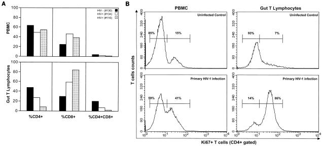
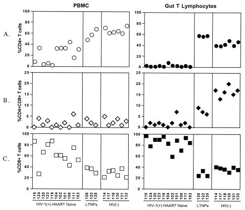
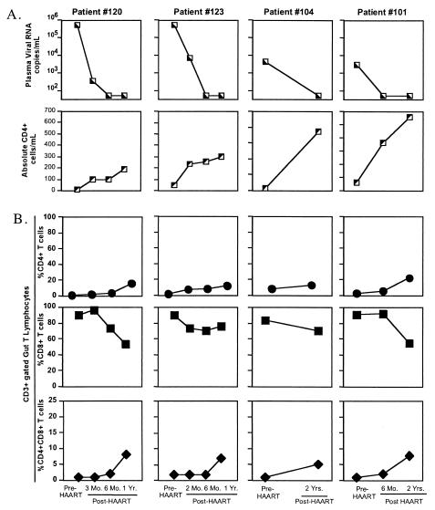
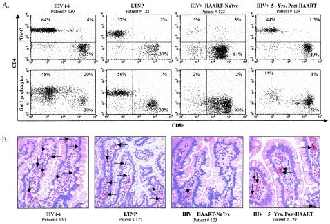
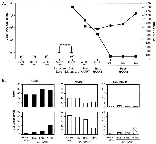
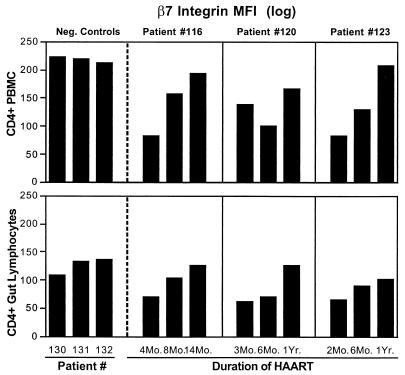
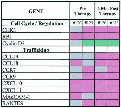
Gut-associated lymphoid tissue (GALT) harbors the majority of T lymphocytes in the body and is an important target for human immunodeficiency virus type 1 (HIV-1). We analyzed longitudinal jejunal biopsy samples from HIV-1-infected patients, during both primary and chronic stages of HIV-1 infection, prior to and following the initiation of highly active antiretroviral therapy (HAART) to determine the onset of CD4(+) T-cell depletion and the effect of HAART on the restoration of CD4(+) T cells in GALT. Severe depletion of intestinal CD4(+) T cells occurred during primary HIV-1 infection. Our results showed that the restoration of intestinal CD4(+) T cells following HAART in chronically HIV-1-infected patients was substantially delayed and incomplete. In contrast, initiation of HAART during early stages of infection resulted in near-complete restoration of intestinal CD4(+) T cells, despite the delay in comparison to peripheral blood CD4(+) T-cell recovery. DNA microarray analysis of gene expression profiles and flow-cytometric analysis of lymphocyte homing and cell proliferation markers demonstrated that cell trafficking to GALT and not local proliferation contributed to CD4(+) T-cell restoration. Evaluation of jejunal biopsy samples from long-term HIV-1-infected nonprogressors showed maintenance of normal CD4(+) T-cell levels in both GALT and peripheral blood. Our results demonstrate that near-complete restoration of mucosal immune system can be achieved by initiating HAART early in HIV-1 infection. Monitoring of the restoration and/or maintenance of CD4(+) T cells in GALT provides a more accurate assessment of the efficacy of antiviral host immune responses as well as HAART.


Figures

FIG. 1.
FIG. 1.
(A) Onset of severe depletion of CD4+ T-cell subsets in GALT occurs in primary HIV-1 infection. Prevalence of CD4+, CD8+, and CD4+CD8+ T-cell subsets in gut T lymphocytes and peripheral blood mononuclear cells (PBMC) of two HAART-naïve individuals (patients 134 and 116) during primary HIV-1 infection was analyzed by flow cytometry and compared with that in uninfected healthy controls. (B) Increased proliferation of intestinal CD4+ T cells in response to primary HIV-1 infection. Flow-cytometric analysis was performed to determine Ki67 expression in CD4+ T cells in peripheral blood and gut T lymphocytes 4 weeks after a suspected high-risk exposure to HIV-1 infection (patient 134), in comparison to a healthy uninfected control.
FIG. 2.
FIG. 2.
Alterations in CD4+ and CD8+ T-cell subsets in GALT of HIV-1-infected individuals with diverse clinical outcomes. Flow-cytometric analysis was performed to determine changes in percentages of CD3-gated CD4+, CD4+CD8+, and CD8+ T cells in gut T lymphocytes and peripheral blood mononuclear cells (PBMC) from HAART-naïve chronically HIV-1-infected individuals (n = 11), LTNPs (n = 3), and uninfected healthy controls (n = 6). Absolute CD4+ T-cell numbers, viral loads, and length of infection at the time of sample collection are presented in Table 1.
FIG. 3.
FIG. 3.
Delayed and incomplete restoration of CD4+ T-cell subsets in GALT compared to peripheral blood in chronically HIV-1-infected patients receiving HAART. (A) Longitudinal analysis of viral loads and absolute CD4+ T-cell numbers in peripheral blood of HIV-1-infected patients (n = 4) prior to and following HAART. (B) Flow-cytometric analysis was performed to determine alterations in percentages of CD3-gated CD4+, CD8+, and CD4+CD8+ T cells in longitudinal jejunal biopsy samples from HIV-1-infected patients prior to and following HAART.
FIG. 4.
FIG. 4.
Flow-cytometric and immunohistochemical analysis of CD4+ T-cell depletion in GALT of HIV-1-infected individuals. (A) Representative dot plots of CD3-gated CD4+ and CD8+ T cells in peripheral blood and GALT; (B) immunohistochemical detection of CD4+ cells of an HIV-negative healthy control, an LTNP, a HAART-naïve chronically HIV-1-infected patient, and an HIV-1-infected patient who had received HAART for 5 years.
FIG. 5.
FIG. 5.
Initiation of HAART during primary HIV-1 infection results in near-complete restoration of CD4+ T cells in GALT. (A) Time line, plasma viral loads, and absolute CD4+ T-cell numbers in peripheral blood of patient 116 during early HIV-1 infection and following the initiation of HAART. ▪, viral RNA copies; •, CD4+ cells. (B) Alterations in percentages of CD4+, CD4+CD8+, and CD8+ T cells in longitudinal jejunal biopsy samples and peripheral blood samples of patient 116, prior to and following HAART. −, HIV-1 seronegative; +, plasma HIV-1 RNA positive.
FIG. 6.
FIG. 6.
Increased expression of lymphocyte homing marker on repopulating CD4+ T cells in GALT following HAART. Flow-cytometric analysis of integrin β7 expression was performed on CD4+ T cells in longitudinal jejunal and peripheral blood samples of three healthy uninfected controls and three HIV-1-infected patients following HAART.
FIG. 7.
FIG. 7.
DNA microarray analysis of gene expression profiles in longitudinal jejunal biopsy samples of HIV-1-infected patients prior to and following HAART. Red indicates increased gene expression compared to five HIV-1-negative controls, green indicates decreased gene expression, and grey indicates no change in the level of gene expression.

References

    1. Ameisen, J. C., and A. Capron. 1991. Cell dysfunction and depletion in AIDS: the programmed cell death hypothesis. Immunol. Today 12:102-105. - PubMed
    1. Andersson, J., T. E. Fehniger, B. K. Patterson, J. Pottage, M. Agnoli, P. Jones, H. Behbahani, and A. Landay. 1998. Early reduction of immune activation in lymphoid tissue following highly active HIV therapy. AIDS 12:F123-F129. - PubMed
    1. Autran, B., G. Carcelain, T. S. Li, C. Blanc, D. Mathez, R. Tubiana, C. Katlama, P. Debre, and J. Leibowitch. 1997. Positive effects of combined antiretroviral therapy on CD4+ T cell homeostasis and function in advanced HIV disease. Science 277:112-116. - PubMed
    1. Brandtzaeg, P. 1989. Overview of the mucosal immune system. Curr. Top. Microbiol. Immunol. 146:13-25. - PubMed
    1. Haase, A. T. 1999. Population biology of HIV-1 infection: viral and CD4+ T cell demographics and dynamics in lymphatic tissues. Annu. Rev. Immunol. 17:625-656. - PubMed

Publication types

MeSH terms