. 2003 Nov;77(21):11754-66.
doi: 10.1128/jvi.77.21.11754-11766.2003.

Evidence against an essential role of COPII-mediated cargo transport to the endoplasmic reticulum-Golgi intermediate compartment in the formation of the primary membrane of vaccinia virus

Affiliations

Evidence against an essential role of COPII-mediated cargo transport to the endoplasmic reticulum-Golgi intermediate compartment in the formation of the primary membrane of vaccinia virus

Matloob Husain et al. J Virol. 2003 Nov.

Abstract

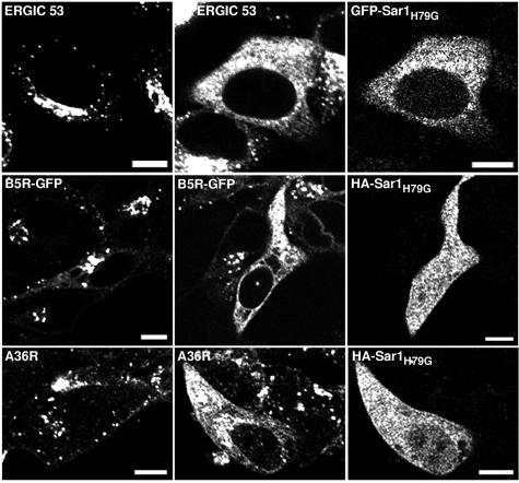
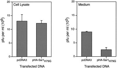
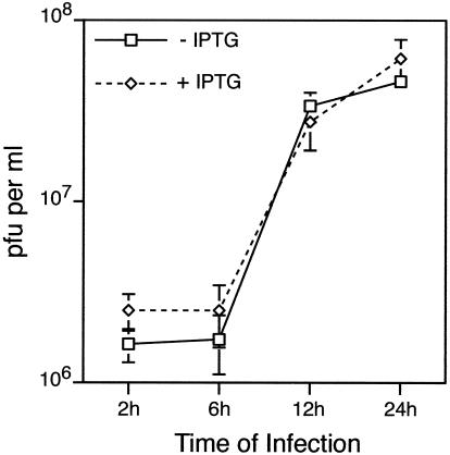
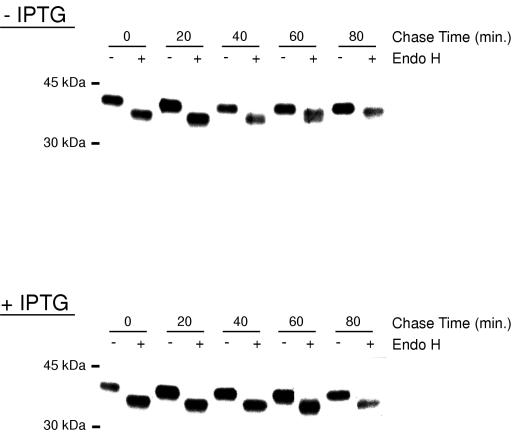
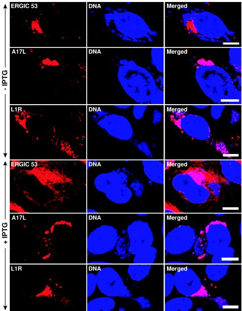
Vaccinia virus assembles two distinct lipoprotein membranes. The primary membrane contains nonglycosylated proteins, appears as crescents in the cytoplasm, and delimits immature and mature intracellular virions. The secondary or wrapping membrane contains glycoproteins, is derived from virus-modified trans-Golgi or endosomal cisternae, forms a loose coat around some intracellular mature virions, and becomes the envelope of extracellular virions. Although the mode of formation of the wrapping membrane is partially understood, we know less about the primary membrane. Recent reports posit that the primary membrane originates from the endoplasmic reticulum-Golgi intermediate compartment (ERGIC). According to this model, viral primary membrane proteins are cotranslationally inserted into the ER and accumulate in the ERGIC. To test the ERGIC model, we employed Sar1(H79G), a dominant negative form of the Sar1 protein, which is an essential component of coatomer protein II (COPII)-mediated cargo transport from the ER to the ERGIC and other post-ER compartments. Overexpression of Sar1(H79G) by transfection or by a novel recombinant vaccinia virus with an inducible Sar1(H79G) gene resulted in retention of ERGIC 53 in the ER but did not interfere with localization of viral primary membrane proteins in factory regions or with formation of viral crescent membranes and infectious intracellular mature virions. Wrapping of intracellular mature virions and formation of extracellular virions did not occur, however, because some proteins that are essential for the secondary membrane were retained in the ER as a consequence of Sar1(H79G) overexpression. Our data argue against an essential role of COPII-mediated cargo transport and the ERGIC in the formation of the viral primary membrane. Instead, viral membranes may be derived directly from the ER or by a novel mechanism.


Figures

FIG. 1.
FIG. 1.
Effect of Sar1H79G expression on the intracellular distribution of cellular ERGIC 53 and vaccinia virus IEV proteins. (Row 1) HeLa cells were either not transfected (column 1) or transfected with pGFP-Sar1H79G (columns 2 and 3) and incubated for 24 h. Cells were fixed, permeabilized, stained with an anti-ERGIC 53 MAb followed by treatment with Alexa 594-conjugated anti-mouse IgG, and viewed by confocal microscopy. (Row 2) HeLa cells were mock transfected (column 1) or transfected (columns 2 and 3) with pHA-Sar1H79G and infected 24 h later with vB5R-GFP (columns 1 to 3). At 24 h after infection, cells were fixed, permeabilized, and stained with anti-HA mouse MAb followed by rhodamine red-conjugated anti-mouse IgG (columns 2 and 3). (Row 3) HeLa cells were mock transfected (column 1) or transfected (columns 2 and 3) with pHA-Sar1H79G and infected 24 h later with vaccinia virus strain WR (columns 1 to 3). At 24 h after infection, cells were treated as described above and stained with anti-A36R rabbit Ab followed by tetramethyl rhodamine isothiocyanate-conjugated anti-rabbit IgG (column 1) or were stained with anti-A36R rabbit Ab and anti-HA mouse MAb followed by tetramethyl rhodamine isothiocyanate-conjugated anti-rabbit IgG and fluorescein isothiocyanate-conjugated anti-mouse IgG (columns 2 and 3). Bars, 10 μm.
FIG.2.
FIG.2.
Effect of Sar1H79G expression on the intracellular distribution of IMV proteins. (Rows 1 and 2) HeLa cells were infected with vA9L-HA (column 1) or WR (columns 2 and 3) and after 24 h were fixed, permeabilized, and stained with anti-HA mouse MAb (column 1), anti-L1R mouse MAb (column 2), or anti-A17LC rabbit Ab (column 3). (Rows 3 to 5) Cells were transfected with pGFP-Sar1H79G (columns 1 and 2) or pHA-Sar1H79G (column 3) and 24 h later were infected as described for rows 1 and 2. Cells were treated as described for rows 1 and 2 except that the cells in column 3 were also stained with anti-HA mouse MAb. The secondary antibodies were either rhodamine red-conjugated anti-mouse IgG or tetramethyl rhodamine isothiocyanate-conjugated anti-rabbit IgG. All cells were counterstained with the DNA binding dye DAPI. Arrows show examples of proteins colocalizing with viral DNA factories. Red fluorescence, viral proteins; blue fluorescence, DNA; green fluorescence, Sar1H79G. Bars, 10 μm.
FIG. 3.
FIG. 3.
Effect of Sar1H79G on the yields of cell-associated and released infectious virus. HeLa cells were transfected with pcDNA3 or pHA-Sar1H79G and infected 24 h later with vaccinia virus strain IHD-J. After a further 24 h, the medium was collected and cleared by high-speed centrifugation, and the cells were harvested in fresh medium, washed, and lysed. Infectious particles were determined by plaque assay on BS-C-1 cells. Data presented are the averages, with standard deviations, of three different plaque counts of two independent experiments.
FIG. 4.
FIG. 4.
Characterization of a recombinant vaccinia virus expressing an inducible GFP-Sar1H79G fusion protein. (A) Diagram of portions of recombinant vaccinia virus vGFP-Sar1H79Gi. Abbreviations: Tk, thymidine kinase gene; T7 pol, bacteriophage T7 polymerase gene; lacO, E. coli lac operator; PL, vaccinia virus P11 late promoter; PE/L, vaccinia virus P7.5 early/late promoter; lacI, E. coli lac repressor gene; HA, vaccinia virus hemagglutinin gene; gpt, E. coli guanine phosphotransferase gene; SLO, stem loop lacO; EMC, encephalomyocarditis virus untranslated leader sequence; GFP-Sar1H79G, ORF encoding mutated GFP-Sar1H79G fusion protein; TT, transcription termination sequences. (B) Immunoblot showing inducible expression of GFP-Sar1H79G. HeLa cells were infected with vGFP-Sar1H79Gi in the absence and presence of 100 μM IPTG for 20 h. Proteins from the cell lysates were resolved by SDS-polyacrylamide gel electrophoresis, transferred to a nitrocellulose membrane, and detected with anti-GFP mouse MAb. (C) Effect of IPTG on plaque size of vGFP-Sar1H79Gi. BS-C-1 cells were infected with vGFP-Sar1H79Gi and overlaid with medium containing methylcellulose and 0 or 100 μM IPTG. After 3 days, plaques were visualized with crystal violet in 20% ethanol. (D) Images of vGFP-Sar1H79Gi plaques with or without IPTG under visible (left) or fluorescent (right) light.
FIG. 5.
FIG. 5.
One-step growth of vGFP-Sar1H79Gi in the presence or absence of IPTG. BS-C-1 cells were infected with 5 PFU of vGFP-Sar1H79Gi per cell in the absence or presence of 100 μM IPTG. Cells were harvested from triplicate cultures at 2, 6, 12, and 24 h after infection, and the infectivity of each was determined by plaque assay. Standard deviations are shown by error bars.
FIG. 6.
FIG. 6.
Effect of IPTG on the intracellular distribution of GFP-Sar1H79G and IEV membrane proteins in vGFP-Sar1H79Gi-infected cells. HeLa cells were infected with vGFP-Sar1H79Gi in the absence (left column) or presence (right column) of IPTG for 20 h. Cells were fixed, permeabilized, and stained with anti-A33R mouse MAb, anti-A36R rabbit Ab, anti-F13L rabbit Ab, and anti-B5R rat MAb, followed by Alexa 594-conjugated anti-mouse IgG, tetramethyl rhodamine isothiocyanate-conjugated anti-rabbit IgG, and anti-rat IgG, respectively. Bars, 10 μm.
FIG. 7.
FIG. 7.
Endo H sensitivity of the B5R glycoprotein synthesized in the absence or presence of IPTG. BS-C-1 cells were infected with 5 PFU of vGFP-Sar1H79Gi per cell in the absence or presence of IPTG. After 8 h, cells were pulse-labeled with [35S]methionine and [35S]cysteine for 5 min and then washed and chased for 0, 20, 40, 60, and 80 min in medium supplemented with unlabeled cysteine, methionine, and IPTG as required. Cells were lysed immediately after the pulse or chase, and the B5R glycoprotein was captured with Ab, subjected to Endo H digestion, resolved by SDS-polyacrylamide gel electrophoresis, and visualized by autoradiography.
FIG. 8.
FIG. 8.
Effect of IPTG on the intracellular location of viral membrane proteins in cells infected with vGFP-Sar1H79Gi. HeLa cells were infected with vGFP-Sar1H79Gi in the absence (rows 1 to 3) or presence (rows 4 to 6) of IPTG for 20 h. The cells were fixed, permeabilized, and stained with anti-ERGIC 53 MAb, anti-L1R mouse MAb, and anti-A17LC rabbit Ab, followed by Alexa 594-conjugated anti-mouse IgG and tetramethyl rhodamine isothiocyanate-conjugated anti-rabbit IgG, respectively. Cells were then stained with DAPI. Red, secondary antibodies; blue, DAPI. Bars, 10 μm.
FIG.9.
FIG.9.
Effect of Sar1H79G expression on the maturation and production of IMV and IEV. RK13 cells were infected with vGFP-SarH79Gi at 5 PFU per cell in the absence or presence of 100 μM IPTG. At 20 after infection, cells were harvested and processed for electron microscopy. All forms of vaccinia virus, including CEV (A), IMV (B), and IEV (C), were visualized in the cells infected without IPTG, whereas only IV (E) and IMV (F) but no CEV or IEV (D) were detected in the cells infected in the presence of IPTG.
FIG. 10.
FIG. 10.
Intracellular localization of ERGIC 53 in cells infected with vaccinia virus strain WR. HeLa cells were infected with WR and at 6, 8, and 24 h after infection were fixed, permeabilized, and stained with mouse anti-ERGIC 53 MAb, followed by Alexa 488-conjugated anti-mouse IgG. Cells were counterstained with DAPI. Green, Alexa 488; blue, DAPI. Note that ERGIC 53 is excluded from viral DNA factories at all time points. Bars, 10 μm.

Similar articles

Cited by

References

    1. Andres, G., R. Garcia-Escudero, C. Simon-Mateo, and E. Vinuela. 1998. African swine fever virus is enveloped by a two-membraned collapsed cisterna derived from the endoplasmic reticulum. J. Virol. 72:8988-9001. - PMC - PubMed
    1. Aridor, M., S. I. Bannykh, T. Rowe, and W. E. Balch. 1995. Sequential coupling between COPII and COPI vesicle coats in endoplasmic reticulum to Golgi transport. J. Cell Biol. 131:875-893. - PMC - PubMed
    1. Aridor, M., K. N. Fish, S. Bannykh, J. Weissman, T. H. Roberts, J. Lippincott-Schwartz, and W. E. Balch. 2001. The Sar1 GTPase coordinates biosynthetic cargo selection with endoplasmic reticulum export site assembly. J. Cell Biol. 152:213-229. - PMC - PubMed
    1. Barlowe, C., L. Orci, T. Yeung, M. Hosobuchi, S. Hamamoto, N. Salama, M. F. Rexach, M. Ravazzola, M. Amherdt, and R. Schekman. 1994. COPII: a membrane coat formed by Sec proteins that drive vesicle budding from the endoplasmic reticulum. Cell 77:895-907. - PubMed
    1. Betakova, T., E. J. Wolffe, and B. Moss. 1999. Regulation of vaccinia virus morphogenesis: phosphorylation of the A14L and A17L membrane proteins and C-terminal truncation of the A17L protein are dependent on the F10L protein kinase. J. Virol. 73:3534-3543. - PMC - PubMed

LinkOut - more resources