. 2003 Nov;77(21):11822-32.
doi: 10.1128/jvi.77.21.11822-11832.2003.

Dengue virus induces novel changes in gene expression of human umbilical vein endothelial cells

Affiliations

Dengue virus induces novel changes in gene expression of human umbilical vein endothelial cells

Rajas V Warke et al. J Virol. 2003 Nov.

Erratum in

  • J Virol. 2004 May;78(9):4947-8

Abstract

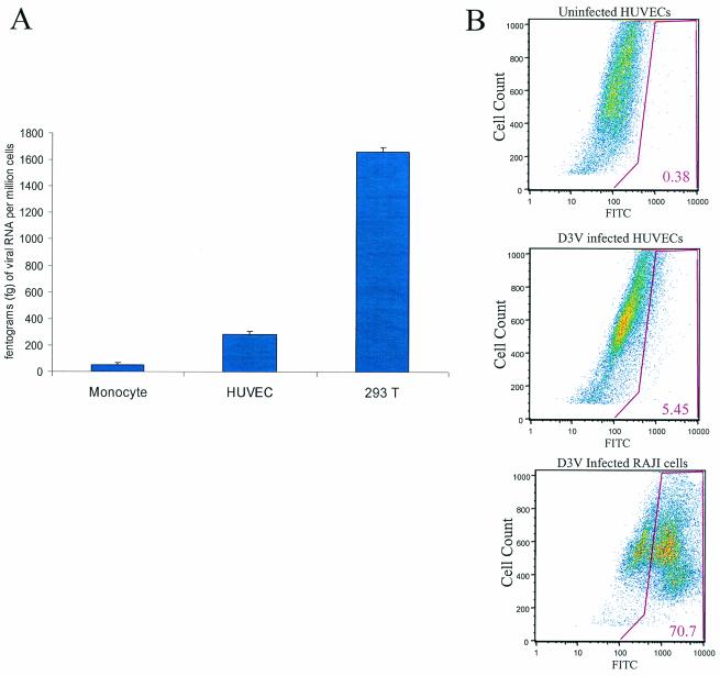
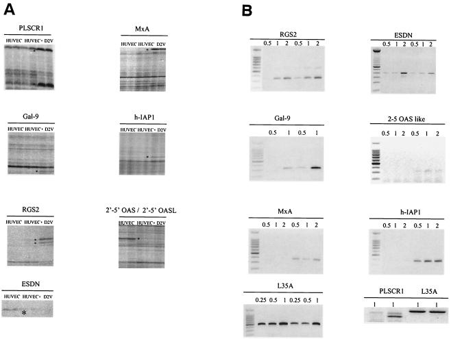
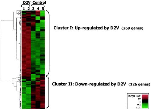
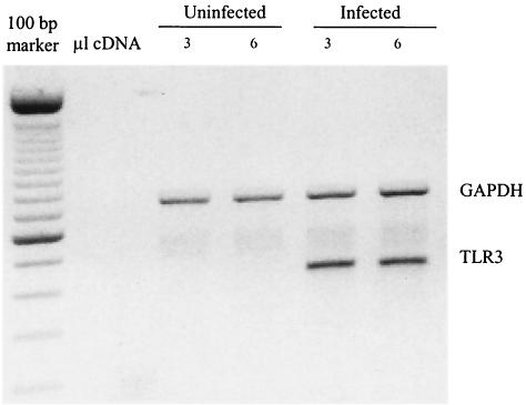
Endothelial cells are permissive to dengue virus (DV) infection in vitro, although their importance as targets of DV infection in vivo remains a subject of debate. To analyze the virus-host interaction, we studied the effect of DV infection on gene expression in human umbilical vein endothelial cells (HUVECs) by using differential display reverse transcription-PCR (DD-RTPCR), quantitative RT-PCR, and Affymetrix oligonucleotide microarrays. DD identified eight differentially expressed cDNAs, including inhibitor of apoptosis-1, 2'-5' oligoadenylate synthetase (OAS), a 2'-5' OAS-like (OASL) gene, galectin-9, myxovirus protein A (MxA), regulator of G-protein signaling, endothelial and smooth muscle cell-derived neuropilin-like protein, and phospholipid scramblase 1. Microarray analysis of 22,000 human genes confirmed these findings and identified an additional 269 genes that were induced and 126 that were repressed more than fourfold after DV infection. Broad functional responses that were activated included the stress, defense, immune, cell adhesion, wounding, inflammatory, and antiviral pathways. These changes in gene expression were seen after infection of HUVECs with either laboratory-adapted virus or with virus isolated directly from plasma of DV-infected patients. Tumor necrosis factor alpha, OASL, and MxA and h-IAP1 genes were induced within the first 8 to 12 h after infection, suggesting a direct effect of DV infection. These global analyses of DV effects on cellular gene expression identify potentially novel mechanisms involved in dengue disease manifestations such as hemostatic disturbance.


Figures

FIG. 1.
FIG. 1.
Primary isolate of DV infects HUVECs in vitro. (A) Quantitative Taqman PCR. We measured DV RNA levels in plasma by quantitative fluorogenic RT-PCR in two subjects with documented D3V viremia. PEG precipitation was used to isolate DV from 120 μl of sera obtained on day 3 of illness (see Materials and Methods). The PEG-precipitated virus was added to three different cell types: primary monocytes isolated from healthy donors, HUVECs, and epithelial human cell line 293T. Cell cultures were infected at an MOI of ∼0.1 for 48 h, and cellular RNA was isolated. The amount of cellular RNA was normalized by actin mRNA levels. Particles of D3V RNA/million cells detected by D3V TaqMan primers and probes (Materials and Methods) were plotted in the three cell types to compare the efficiencies of infection. The measurements were done in triplicate, and the standard error is shown for each cell type. The results from a representative patient are shown. (B) Indirect immunofluorescence. The percentage of infection of HUVECs with clinical isolates of D3V was detemined by flow cytometry. HUVECs were infected with PEG-precipitated D3V for 48 h, and the cells were fixed by using 1% formaldehyde. The cells were stained with mouse anti-D3V antibody.
FIG. 2.
FIG. 2.
(A) Differentially displayed genes in HUVEC 48 h after D2V infection. HUVEC RNA was used to prepare cDNA with SensiScript reverse transcriptase and HT11C primer. Asterisks indicate differentially displayed bands identified in DV-infected cultures (D2V) compared to uninfected cells (HUVEC). +, addition of exogenous viral RNA to uninfected cells. (B) Semiquantitative RT-PCR analysis. One of three semiquantitative RT-PCR analyses is shown for seven of the eight genes identified by DD; uninfected (left) or virus-infected cultures (right) at 48 h postinfection are included in each panel. L35A was used as a control gene. We used 0.5, 1, and 2 μl of cDNA of either uninfected and infected samples in the PCR for all reactions except for L35A, in which 0.25, 0.5, and 1 μl of cDNA were used. The PLSCR1 gene analysis was performed independently. For the PLSCR1 RT-PCR, results obtained with 1 μl of cDNA are shown. Panel B, left side, uninfected cells; right side, infected cells. The PLSCR1 panel has uninfected cells in lanes 1 and 3 and infected cells in lanes 2 and 4.
FIG. 3.
FIG. 3.
Affymetrix H133A analysis. Hierarchical cluster analysis of 22,283 human genes measured by using Affymetrix gene chips. The results were filtered to retain only the 395 genes differentially expressed by >4-fold. Cluster I (269 genes) was upregulated, and cluster II (126 genes) was downregulated by infection. The key shows the range of relative differential expression of the 395 genes. Displayed expression levels were standardized. A full cluster diagram with all genes identities is available at our website at http://biotools.umassmed.edu/ibosch/.
FIG. 4.
FIG. 4.
Multiplex PCR for the expression of TLR1 to -5 in uninfected and D2V-infected HUVECs. cDNA template was obtained from an RT step performed on total RNA isolated from control and D2V-infected cultures 48 h postinfection. GAPDH (glyceraldehyde-3-phosphate dehydrogenase) is given as a control for equal loading. In the PCR step, two different quantities of cDNA (3 and 6 μl) were used. GAPDH is shown at 680 bp.

References

    1. Alexopoulou, L., A. C. Holt, R. Medzhitov, and R. A. Flavell. 2001. Recognition of double-stranded RNA and activation of NF-κB by Toll-like receptor 3. Nature 413:732-738. - PubMed
    1. Anderson, R., S. Wang, W. Osiowy, and A. Iseekuts. 1997. Activation of endothelial cells via antibody-enhanced dengue virus infection of peripheral blood monocytes. J. Immunol. 71:4226-4434. - PMC - PubMed
    1. Avirutnan, P., P. Malasit, B. Seliger, S. Bhakdi, and M. Husmann. 1998. Dengue virus infection of human endothelial cells leads to chemokine production, complement activation, and apoptosis. J. Immunol. 161:6338-6346. - PubMed
    1. Barondes, S. H., V. Castronovo, D. N. Cooper, R. D. Cummings, K. Drickamer, T. Feizi, M. A. Gitt, J. Hirabayashi, C. Hughes, K. Kasai, et al. 1994. Galectins: a family of animal β-galactoside-binding lectins. Cell 76:597-598. - PubMed
    1. Bonner, S. M., and M. A. O'Sullivan. 1998. Endothelial cell monolayers as a model system to investigate dengue shock syndrome. J. Virol. Methods 71:159-167. - PubMed

Publication types