. 2003 Oct 28;100(22):12543-7.
doi: 10.1073/pnas.2035257100. Epub 2003 Oct 14.

DNA-mediated charge transport for DNA repair

Affiliations

DNA-mediated charge transport for DNA repair

Elizabeth M Boon et al. Proc Natl Acad Sci U S A. .

Erratum in

  • Proc Natl Acad Sci U S A. 2004 Mar 30;101(13):4718

Abstract

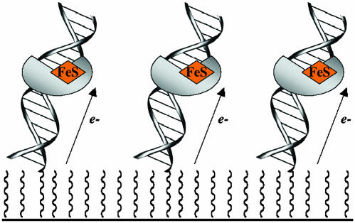
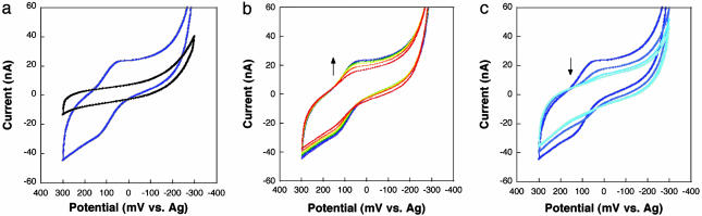
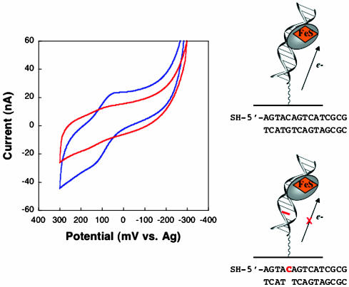
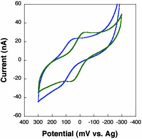
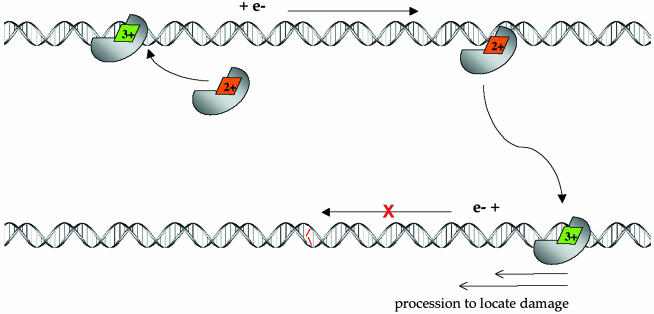
MutY, like many DNA base excision repair enzymes, contains a [4Fe4S]2+ cluster of undetermined function. Electrochemical studies of MutY bound to a DNA-modified gold electrode demonstrate that the [4Fe4S] cluster of MutY can be accessed in a DNA-mediated redox reaction. Although not detectable without DNA, the redox potential of DNA-bound MutY is approximately 275 mV versus NHE, which is characteristic of HiPiP iron proteins. Binding to DNA is thus associated with a change in [4Fe4S]3+/2+ potential, activating the cluster toward oxidation. Given that DNA charge transport chemistry is exquisitely sensitive to perturbations in base pair structure, such as mismatches, we propose that this redox process of MutY bound to DNA exploits DNA charge transport and provides a DNA signaling mechanism to scan for mismatches and lesions in vivo.


Figures

Fig. 1.
Fig. 1.
Schematic illustration of DNA-modified electrodes used in these studies to determine the DNA-bound redox potential of MutY electrochemically.
Fig. 2.
Fig. 2.
CV of 800 μM MutY at a gold surface modified with the thiol-terminated duplex SH-5′-AGTACAGTCATCGCG and passivated with mercaptohexanol. (a) The redox potential of MutY measured in these experiments requires DNA binding. The CV of MutY at a surface modified with mercaptohexanol (MCH) only (black) shows no redox process, whereas at a DNA/MCH-modified Au surface (blue), the DNA-bound redox potential of MutY is ≈75 mV vs. Ag (≈275 mV vs. NHE). (b and c) This electrochemical signal grows in with DNA-binding time (CVs for incubation from 1–10 min) (b) and persists as the MutY solution is exchanged for pure buffer (20 mM sodium phosphate, pH 7.5/1 mM Na2EDTA/20% glycerol/500 mM NaCl) (c), indicating that MutY is bound to the DNA-modified surface.
Fig. 3.
Fig. 3.
The redox chemistry of MutY is DNA-mediated. CV of 800 μM MutY at a surface modified with the thiol-terminated duplex SH-5′-AGTACAGTCATCGCG hybridized to a fully base-paired complement (blue) versus a complement with an abasic site opposite the red C is shown.
Fig. 4.
Fig. 4.
CV of 800 μM WT (blue) and C199H mutant (green) MutY at a gold electrode modified with the thiol-terminated sequence SH-5′-AGTACAGTCATCGCG hybridized to a fully base-paired complement. A –70 mV potential shift is observed with the cluster mutant.
Fig. 5.
Fig. 5.
Schematic model of long-range scanning for mismatches by MutY through DNA-mediated CT chemistry. (Upper) Nonspecific binding of MutY to DNA, where binding is associated with a shift in redox potential of the [4Fe4S]2+ cluster, leading to oxidation to the 3+ form. Associated with cluster oxidation is DNA-mediated CT to an alternately bound MutY, where reduction to the 2+ cluster promotes dissociation of the protein. Because this CT process proceeds without interruption by an intervening mismatch, the process constitutes a scan of this region of the genome. (Lower) Association of MutY to a region containing a mismatch (red), where the associated stacking perturbation does not permit DNA-mediated CT to occur. Here, the protein is shown processively diffusing to the mismatch site.

References

    1. Guan, Y., Manuel, R. C., Arvai, A. S., Parikh, S. S., Mol, C. D., Miller, J. H., Lloyd, S. & Tainer, J. A. (1998) Nat. Struct. Biol. 5 1058–1064. - PubMed
    1. Cunningham, R. P., Asahara, H., Bank, J. F., Scholes, C. P., Salerno, J. C., Surerus, K., Munck, E., McCracken, J., Peisach, J. & Emptage, M. H. (1989) Biochemistry 28 4450–4455. - PubMed
    1. Pomposiello, P. J. & Demple, B. (2001) Trends Biotechnol. 19 109–114. - PubMed
    1. Unden, G. & Bongaerts, J. (1997) Biochim. Biophys. Acta 1320 217–222. - PubMed
    1. Rebeil, R., Sun, Y., Chooback, L., Pedraza-Reyes, M., Kinsland, C., Begley, T. P. & Nicholson, W. L. (1998) J. Bacteriol. 180 4879–4885. - PMC - PubMed

Publication types

LinkOut - more resources