

doi: 10.1073/pnas.2135496100.
Epub 2003 Oct 14.
Principles of cell-free genetic circuit assembly
Affiliations
- PMID: 14559971
- PMCID: PMC240676
- DOI: 10.1073/pnas.2135496100
Item in Clipboard
Principles of cell-free genetic circuit assembly
Proc Natl Acad Sci U S A.
.
Abstract
Cell-free genetic circuit elements were constructed in a transcription-translation extract. We engineered transcriptional activation and repression cascades, in which the protein product of each stage is the input required to drive or block the following stage. Although we can find regions of linear response for single stages, cascading to subsequent stages requires working in nonlinear regimes. Substantial time delays and dramatic decreases in output production are incurred with each additional stage because of a bottleneck at the translation machinery. Faster turnover of RNA message can relieve competition between genes and stabilize output against variations in input and parameters.
Figures
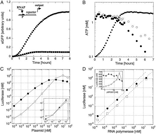
One-stage network. (A) A reporter gene (luc or egfp, promoter P) is expressed in the coupled transcription/translation T7/SP6 RNA polymerase wheat germ extract. Kinetics of expression of EGFP in LLM extract (0.1 nM SP6-egfp, filled circles) and SLM extract (0.5 nM T7-egfp, filled squares) with 20 nM T7 RNA polymerase. Three phases are observed: a lag phase of 10 min, an accumulation of synthesized EGFP, and a slowing down to a plateau. The initial lag phase is for transcription and translation. In the SLM extract, expression stops after 3 h, whereas, in the LLM extract, EGFP keeps accumulating beyond 6 h. (B) ATP concentration measurement in the SLM extract. With 20 nM T7 RNA polymerase and 0.5 nM T7-eGFP plasmid (filled circles) and without expression (open circles). The kinetics of expression of EGFP in SLM extract from A has been superimposed (gray filled squares). (C) Luc production in LLM extract measured after 6 h as a function of T7-luc plasmid concentration (filled circles) and SP6-Luc (open squares) with 20 nM RNA polymerase. (Inset) endogenous expression from the extract without addition of T7 or SP6 RNA polymerase as a function of the T7-luc (filled circles) and SP6-luc (open squares) plasmids. (D) Luc production in LLM extract as a function of T7 or SP6 RNA polymerase at 0.1 nM plasmid concentration, T7-luc (filled circles), and SP6-luc (open squares). (Inset) Luc production in LLM extract as a function of neutral plasmid pBR322 concentration, 20 nM RNA polymerase, 0.1 nM T7-luc plasmid (filled circles), and 0.1 nM SP6-luc (open squares).
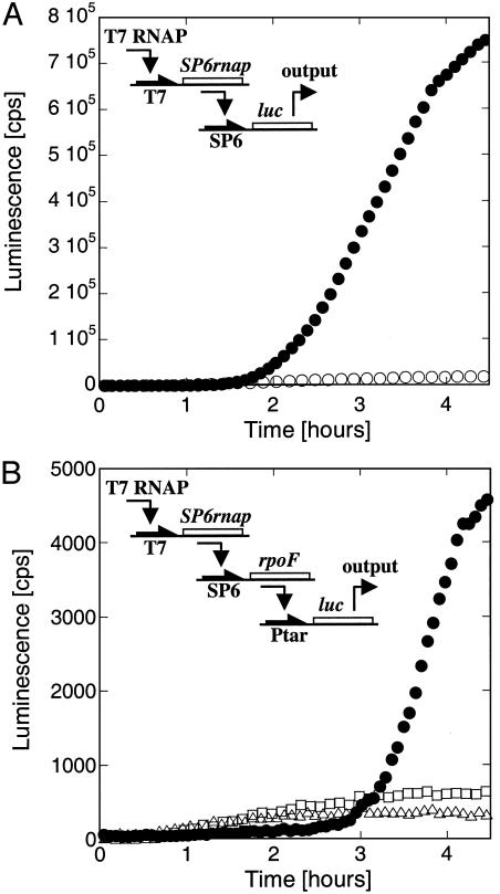
Two and three-stage networks in LLM extract. (A) The SP6 RNA polymerase gene is transcribed by the T7 RNA polymerase. The synthesized SP6 RNA polymerase transcribes the luc gene. Kinetics of expression of the cascade with 20 nM input, 0.5 plasmid T7-SP6rnap, and 2 nM plasmid SP6-luc (filled circles), or 2 nM SP6-luc plasmid only (open circles). (B) The SP6 RNA polymerase gene is transcribed by the T7 RNA polymerase. The SP6 RNA polymerase transcribes the rpoF gene (E. coli sigma factor F), and the synthesized sigma F interacts with the E. coli core enzyme (added to the extract) to induce the transcription of luc gene. To test the kinetics of Luc synthesis: the three-step transcriptional cascade introduces a time delay of 3 h in Luc synthesis (filled circles: 5 nM T7-SP6rnap, 5 nM SP6-rpoF, and 0.5 nM Ptar-luc plasmids); as a control, in the absence of the first or the first two stages, the luc gene is transcribed from the leak of both the T7 RNA polymerase and the E. coli RNA polymerase core enzyme (open triangles, 0.5 nM Ptar-luc plasmid; open squares, 5 nM SP6-rpoF and 0.5 nM Ptar-luc plasmids).
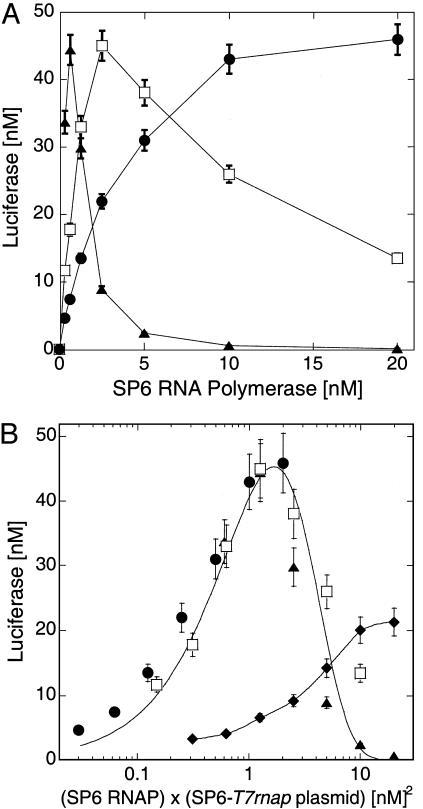
Two-stage network, sharing of resources. The T7 RNA polymerase gene is transcribed by SP6 RNA polymerase. The synthesized T7 RNA polymerase transcribes the luc gene in LLM extract. Expression of the cascade is measured after 6 h. (A) Luc output as a function of SP6 RNA polymerase input in LLM extract with 1 nM T7-luc plasmid and different concentrations of SP6-T7rnap plasmid: 0.1 nM (filled circles), 0.5 nM (open squares), 2 nM (filled triangles). (B) Input–output curves of A (in LLM extract) collapse onto one curve when plotting data as a function of (SP6-T7rnap plasmid) × (SP6 polymerase) (symbols as in A). A monotonous response is observed in SLM extract (filled diamonds), 1 nM SP6-T7rnap and T7-luc plasmid. Solid lines are smoothing fits.
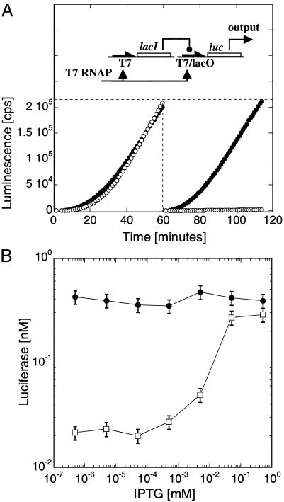
T7/lacO LacI repression. (A) E. coli LacI repressor under the T7 promoter represses the plasmid T7/lacO-luc [pET21(+)-luc] by binding its recognition site, lacO, located downstream of the T7 promoter. Kinetics of coexpression of T7-lacI with T7-luc (filled circles) or T7/lacO-luc (open circles), without (Left) or with (Right) 1 h of preincubation of the plasmid T7-lacI, expression in LLM extract with 0.1 nM of each plasmid. (B) Induction of Luc synthesis as a function of IPTG concentration after 1 h of preincubation of T7-lacI plasmid, followed by 1 h of coexpression with T7/lacO-luc (open squares) or T7-luc (filled circles) (0.5 nM T7-lacI plasmid and 0.1 nM of either T7-luc or T7/lacO-luc plasmids).
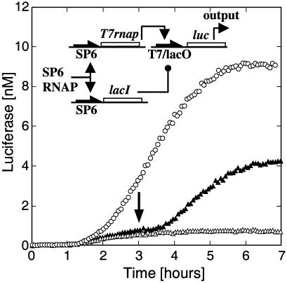
Three-gene circuit. A three-gene circuit with plasmids SP6-lacI, SP6-T7rnap, and T7/lacO-luc (pIVEX2.3d-lacO-luc). Coexpression of the three genes with (open circles) and without (open triangles) IPTG at t = 0 (expression in LLM extract, 0.005 nM SP6-T7rnap, 0.1 nM SP6-lacI and 0.5 nM T7/lacO-luc plasmids). IPTG (0.5 mM) was added after3hto induce Luc production (arrow, filled triangles).
References
Publication types
MeSH terms
Substances
LinkOut - more resources
Full Text Sources
Other Literature Sources
