. 2003 Nov;23(21):7448-59.
doi: 10.1128/MCB.23.21.7448-7459.2003.

XBP-1 regulates a subset of endoplasmic reticulum resident chaperone genes in the unfolded protein response

Affiliations

XBP-1 regulates a subset of endoplasmic reticulum resident chaperone genes in the unfolded protein response

Ann-Hwee Lee et al. Mol Cell Biol. 2003 Nov.

Abstract

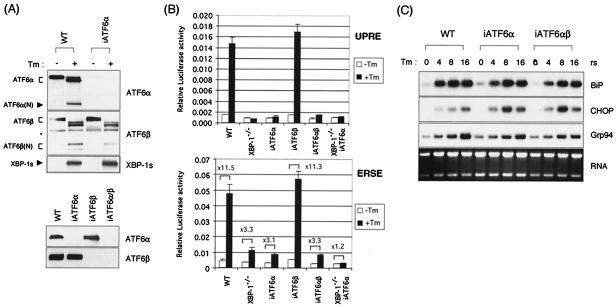
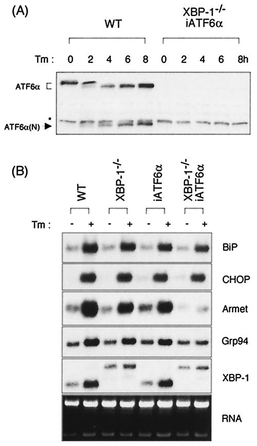
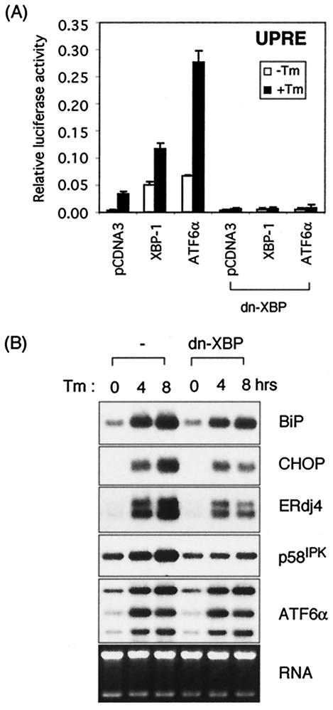
The mammalian unfolded protein response (UPR) protects the cell against the stress of misfolded proteins in the endoplasmic reticulum (ER). We have investigated here the contribution of the UPR transcription factors XBP-1, ATF6alpha, and ATF6beta to UPR target gene expression. Gene profiling of cell lines lacking these factors yielded several XBP-1-dependent UPR target genes, all of which appear to act in the ER. These included the DnaJ/Hsp40-like genes, p58(IPK), ERdj4, and HEDJ, as well as EDEM, protein disulfide isomerase-P5, and ribosome-associated membrane protein 4 (RAMP4), whereas expression of BiP was only modestly dependent on XBP-1. Surprisingly, given previous reports that enforced expression of ATF6alpha induced a subset of UPR target genes, cells deficient in ATF6alpha, ATF6beta, or both had minimal defects in upregulating UPR target genes by gene profiling analysis, suggesting the presence of compensatory mechanism(s) for ATF6 in the UPR. Since cells lacking both XBP-1 and ATF6alpha had significantly impaired induction of select UPR target genes and ERSE reporter activation, XBP-1 and ATF6alpha may serve partially redundant functions. No UPR target genes that required ATF6beta were identified, nor, in contrast to XBP-1 and ATF6alpha, did the activity of the UPRE or ERSE promoters require ATF6beta, suggesting a minor role for it during the UPR. Collectively, these results suggest that the IRE1/XBP-1 pathway is required for efficient protein folding, maturation, and degradation in the ER and imply the existence of subsets of UPR target genes as defined by their dependence on XBP-1. Further, our observations suggest the existence of additional, as-yet-unknown, key regulators of the UPR.


Figures

FIG. 1.
FIG. 1.
Structure of the XBP-1 gene and protein in wt and mutant cells. (A) XBP-1 locus in wt and XBP-1−/− MEF cells. Splicing of the mutant XBP-1 mRNA in XBP-1−/− cells is shown. Asterisks represent termination codons. (B) wt and XBP-1−/− MEF cells were either not treated or treated with 10 μg of Tm/ml for 6 h. XBP-1 mRNA was revealed by Northern blot analysis. (C) wt and XBP-1−/− MEF cells were treated with 10 μM MG-132 or 10 μg of Tm/ml for 6 h. XBP-1u and XBP-1s proteins were detected by Western blot analysis with anti-XBP-1 antibody.
FIG. 2.
FIG. 2.
Dependence of UPR target gene expression on XBP-1. (A) wt and XBP-1−/− MEF cells were treated with 10 μg of Tm/ml for the indicated time periods. Total RNAs were isolated and subjected to Northern blot analysis. The same blot was hybridized sequentially with BiP, CHOP, MGP, ERdj4, p58IPK, ATF6α, and MGP probes. The p58IPK probe is probe A, from the 3′ end of the gene. Ethidium bromide staining of the gel before blotting is shown at the bottom for loading control. (B) All isoforms of p58IPK are XBP-1 dependent. Here, Northern blot analysis was performed with probe B, which recognizes sequences at the 5′ end of the gene. (C) XBP-1-dependent genes are also IRE1α dependent. Northern blot analysis of RNA prepared from IRE1α−/− MEFs treated with Tm for various time periods and assessed for expression of ERdj4 and p58IPK. (D) The ERdj4GL3 reporter was transfected with or without XBP-1s plasmid into wt and XBP-1−/− MEF cells. Cells were treated with Tm at 1 μg/ml for 16 h before harvesting as indicated. Luciferase activity was normalized to the Renilla activity. (E) Induction of BiP, ERdj4, and p58IPK in primary B cells by LPS. B220+ primary B cells were isolated from spleens of wt or XBP-1−/− RAG2−/− lymphoid chimeras. Cells were untreated or stimulated for 3 days with 20 μg of LPS/ml. Expression of BiP, ERdj4, and p58IPK was determined by Northern blot analysis.
FIG. 3.
FIG. 3.
Induction of UPR target genes by XBP-1s. MEF-tet-off and MEF-tet-off-XBP-1s cells were cultured in medium containing 1 μg of doxycycline/ml. XBP-1s expression was induced by culturing the cells for 3 days in doxycycline-free medium or by treatment with Tm for the indicated times. XBP-1s protein was revealed by anti-XBP-1 antibody in Western blot analyses. Total RNA was also prepared to measure the expression level of BiP, CHOP, ERdj4, p58IPK, and ATF6α mRNA. (B) Additional XBP-1-dependent target genes identified in gene profiling experiments (Table 2) were confirmed by Northern analysis. Ethidium bromide staining of the gels before blotting is shown at the bottom.
FIG. 4.
FIG. 4.
UPR target gene expression is largely unaffected in the absence of ATF6α and ATF6β. (A) Western blot analysis of iATF6α-, iATF6β-, and double iATF6α/β-expressing MEFs. MEF-iATF6α cells were generated by transfecting MEF cells with U6-iATF6α plasmid, which expresses siRNA for ATF6α under the control of the U6 promoter. MEF-iATF6β and MEF-iATF6α/β cells were generated by transducing wt and MEF-iATF6α cells with retroviruses that express iATF6β. Lysates from wt and MEF-iATF6α, MEF-iATF6β, and MEF-iATF6α/β MEFs either not treated or treated with 10 μg of Tm/ml for 6 h were analyzed for the expression of XBP-1, ATF6α, and ATF6β. An asterisk indicates a nonspecific band recognized by anti-ATF6α antibody. (B) 5xATF6GL3 (UPRE) or ERSE reporters were transfected into wt, XBP-1−/−, iATF6α, iATF6β, double iATF6α/β, and double XBP-1−/−/iATF6α MEF cells. Cells were treated with Tm at 1 μg/ml for 16 h before harvesting as indicated. Luciferase activity was normalized to Renilla activity. The fold induction of relative luciferase activity by Tm treatment compared to untreated samples is also shown. (C) Total RNA was prepared to measure the expression level of BiP, CHOP, ERdj4, p58IPK, and Grp94 mRNAs. Ethidium bromide staining of the gel before blotting is shown at the bottom.
FIG. 5.
FIG. 5.
UPR target gene expression in cells that lack both XBP-1 and ATF6α. (A) An XBP-1−/−/ATF6α doubly deficient MEF cell line was generated by transferring siRNA for ATF6α into XBP-1−/− MEF cells. ATF6α protein was absent in the doubly deficient cells as confirmed by Western blot analysis with anti-ATF6α antibody. (B) Total RNA was isolated from the indicated cell lines that were untreated or treated with Tm for 6 h and subjected to Northern blot analysis. The same blot was hybridized sequentially with BiP, CHOP, Armet, ERdj4, p58IPK, and Grp94 probes. Ethidium bromide staining of the gel before blotting is shown at the bottom as a loading control.
FIG. 6.
FIG. 6.
Dominant negative XBP-1 suppresses both XBP-1 and ATF6α activity. (A) The 5xATF6GL3 reporter plasmid was cotransfected with either pCGNATF6α or XBP-u/s plasmids into MEF cells with or without the dominant-negative XBP-1 expression plasmid. A total of 100 ng of DNA was used for each transfection except for pCDNA3.1, which was added to give 1 μg of DNA in total. Luciferase assays were performed as described in the legend to Fig. 4. (B) MEF and MEF-dn-XBP cells that stably express dominant-negative XBP-1 protein were treated with 10 μg of Tm/ml for the indicated time periods. Total RNAs were isolated and subjected to Northern blot analysis. The same blot was hybridized sequentially with BiP, CHOP, ERdj4, p58IPK, and ATF6α probes. Ethidium bromide staining of the gel before blotting is shown at the bottom as a loading control.

References

    1. Bukau, B., and A. L. Horwich. 1998. The Hsp70 and Hsp60 chaperone machines. Cell 92:351-366. - PubMed
    1. Calfon, M., H. Zeng, F. Urano, J. H. Till, S. R. Hubbard, H. P. Harding, S. G. Clark, and D. Ron. 2002. IRE1 couples endoplasmic reticulum load to secretory capacity by processing the XBP-1 mRNA. Nature 415:92-96. - PubMed
    1. Chevalier, M., H. Rhee, E. C. Elguindi, and S. Y. Blond. 2000. Interaction of murine BiP/GRP78 with the DnaJ homologue MTJ1. J. Biol. Chem. 275:19620-19627. - PMC - PubMed
    1. Clauss, I. M., M. Chu, J.-L. Zhao, and L. H. Glimcher. 1996. The basic domain/leucine zipper protein hXBP-1 preferentially binds to and transactivates CRE-like sequences containing an ACGT core. Nucleic Acids Res. 24:1855-1864. - PMC - PubMed
    1. Cox, J. S., and P. Walter. 1996. A novel mechanism for regulating activity of a transcription factor that controls the unfolded protein response. Cell 87:391-404. - PubMed

Publication types

MeSH terms