. 2003 Nov;23(21):7460-74.
doi: 10.1128/MCB.23.21.7460-7474.2003.

PU.1 and pRB interact and cooperate to repress GATA-1 and block erythroid differentiation

Affiliations

PU.1 and pRB interact and cooperate to repress GATA-1 and block erythroid differentiation

Natasha Rekhtman et al. Mol Cell Biol. 2003 Nov.

Abstract

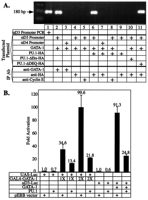
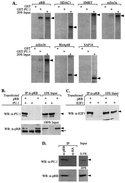
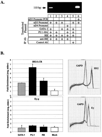
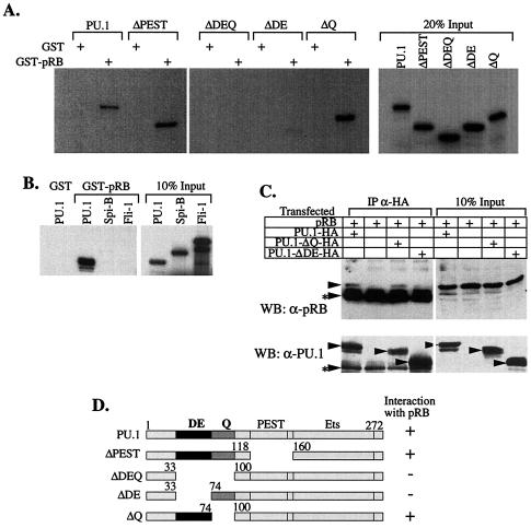
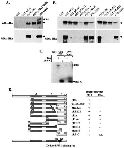
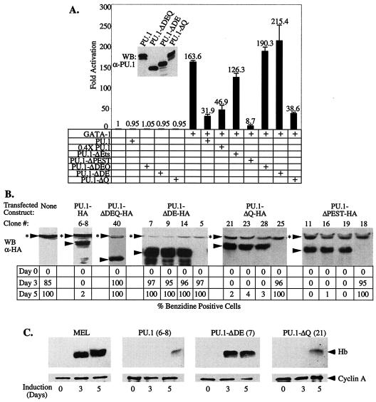
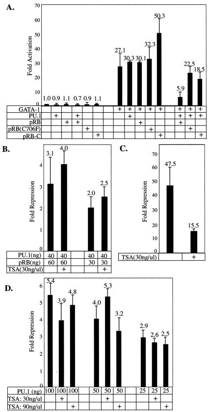
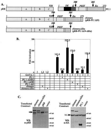
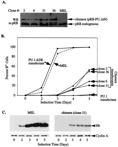
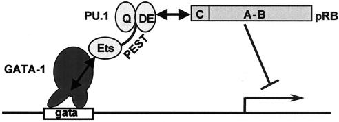
PU.1 and GATA-1 are two hematopoietic specific transcription factors that play key roles in development of the myeloid and erythroid lineages, respectively. The two proteins bind to one another and inhibit each other's function in transcriptional activation and promotion of their respective differentiation programs. This mutual antagonism may be an important aspect of lineage commitment decisions. PU.1 can also act as an oncoprotein since deregulated expression of PU.1 in erythroid precursors causes erythroleukemias in mice. Studies of cultured mouse erythroleukemia cell lines indicate that one aspect of PU.1 function in erythroleukemogenesis is its ability to block erythroid differentiation by repressing GATA-1 (N. Rekhtman, F. Radparvar, T. Evans, and A. I. Skoultchi, Genes Dev. 13:1398-1411, 1999). We have investigated the mechanism of PU.1-mediated repression of GATA-1. We report here that PU.1 binds to GATA-1 on DNA. We localized the repression activity of PU.1 to a small acidic N-terminal domain that interacts with the C pocket of pRB, a well-known transcriptional corepressor. Repression of GATA-1 by PU.1 requires pRB, and pRB colocalizes with PU.1 and GATA-1 at repressed GATA-1 target genes. PU.1 and pRB also cooperate to block erythroid differentiation. Our results suggest that one of the mechanisms by which PU.1 antagonizes GATA-1 is by binding to it at GATA-1 target genes and tethering to these sites a corepressor that blocks transcriptional activity and thereby erythroid differentiation.


Figures

FIG. 1.
FIG. 1.
PU.1 binds to GATA-1 on DNA. (A) A total of 107 U2OS cells were transfected by the Lipofectamine Plus method (Gibco-BRL) with 12.9 μg of DNA consisting of 420 ng of αD3-Luc, 2.8 μg of pXM-GATA-1, 2.8 μg of the indicated pEBB-PU.1-HA expression plasmid, and the required amount of pBSK vector DNA. ChIPs were carried out as described in Materials and Methods with anti-HA, anti-GATA-1, or anti-cyclin E antibodies. The presence of DNA sequences encompassing the GATA-1 site in the αD3 promoter (or the mutant GATA-1 site in the αD4 promoter) was determined by PCR assays as described in Materials and Methods. The presence of the 180-bp fragment indicates presence of the promoter region in the immunoprecipitate. Lane 1 shows PCR amplification of αD3-Luc plasmid DNA. A schematic diagram of the mutant PU.1 proteins used is shown in Fig. 3. (B) U2OS cells were transfected with a UAS-Luc reporter (100 ng) or the αD3-Luc reporter (15 ng), and pRSV-GATA-1-GAL4(DBD) (50 and 100 ng), pXM-GATA-1 (50 ng), and 30 ng of pEBB-PU.1 or 30 ng of empty pEBB vector as indicated. Luciferase activity was determined 48 h after transfection.
FIG. 2.
FIG. 2.
PU.1 and pRB interact in vitro and in vivo. (A) 35S-labeled proteins, indicated above each panel, were prepared by coupled transcription-translation reactions and tested for interaction with GST or GST-PU.1 immobilized on glutathione-Sepharose beads as described in Materials and Methods. Bound proteins were analyzed by sodium dodecyl sulfate-polyacrylamide gel electrophoresis (SDS-PAGE) and autoradiography. An autoradiogram of 20% of each 35S-labeled protein added to the binding reactions is shown in each panel. Arrowheads indicate the positions of the 35S-labeled proteins on the gel. (B and C) Extracts of 293 cells, transfected with expression constructs for PU.1, E2F1, and pRB (large pocket, residues 379 to 928) as indicated, were immunoprecipitated (IP) with anti-pRB (XZ91; Pharmingen) antibody. Immunoprecipitated complexes were resolved by SDS-PAGE and analyzed by Western blotting (WB) with the indicated primary antibodies. Arrowheads mark the position of specifically immunodetected proteins; arrowheads with an asterisk indicate the position of the immunoglobulin chains used in the immunoprecipitaions. An immunoblot of the indicated fraction of each type of extract added to the immunoprecipitation reactions is shown in each panel (% input lanes). (D) Extracts of MEL cells were immunoprecipitated with anti-pRB (G3-245; Pharmingen) antibody and analyzed as in panel B.
FIG. 3.
FIG. 3.
PU.1 and pRB colocalize with GATA-1 at GATA-1 binding sites in chromatin. (A) U2OS cells were transfected as described in the legend to Fig. 1A, and ChIP was carried out as described in Materials and Methods with anti-pRB antibody or an anti-rabbit immunoglobulin G antibody that has the same isotype (Control Ab). PCR assays were performed as described in Materials and Methods with specific primers that differ from the primers used in Fig. 1A; here the presence of the 110-bp fragment indicates the presence of the αD3 promoter region in the immunoprecipitate. Lane 6 shows PCR amplification of αD3-Luc plasmid DNA with the primers. (B) ChIP was performed on cross-linked chromatin from MEL cells as described in Materials and Methods with antibodies to GATA-1, PU.1, and pRB (RB) and with β-tubulin (Mock). The amounts of specific promoter DNA fragments relative to G6PD DNA in duplicate immunoprecipitates were quantitated by real-time PCRs, and the degree of enrichment of the promoter fragments was calculated. Error bars indicate the standard deviation. Panels on the right show the dissociation curves of the set of amplified DNA fragments for HS2 (Tm = 85°C), G6PD (Tm = 78.5°C), and Ey (Tm = 82°C).
FIG. 4.
FIG. 4.
The acidic subdomain of PU.1 is required for interaction of PU.1 with pRB in vitro and in vivo. (A and B) 35S-labled proteins prepared by coupled transcription-translation reactions were tested for interaction with GST or GST-pRB as described in Fig. 2. (C) 293 cells were transfected with pRB (large pocket, residues 379 to 928) and expression vectors encoding PU.1 or PU.1-mutant proteins tagged with an HA epitope. Cell extracts were prepared and immunoprecipitated (IP) with anti-HA antibody. Bound complexes were analyzed by Western blotting with anti-pRB (XZ55) or anti-PU.1 primary antibodies. Other details are as in Fig. 2. (D) Schematic diagram of the wild-type and mutant PU.1 proteins used in the interaction studies in panels A to C and a summary of the results for PU.1 interaction with pRB. DE and Q indicate the acidic and glutamine residue-rich subdomains, respectively; PEST, region rich in PEST residues; Ets, DNA-binding Ets homology domain.
FIG. 5.
FIG. 5.
The C pocket of pRB mediates interaction with PU.1. (A) An expression construct encoding PU.1-HA was transfected into 293 cells, which endogenously express E1A, and cell extracts were tested for interaction with GST or the GST fusion proteins indicated above the panel. Bound proteins were analyzed by SDS-PAGE and Western blot analysis (WB) with anti-HA or anti-E1A primary antibodies. Arrowheads mark the position of the specifically immunodetected proteins. n.s., Position of a nonspecific band detected in 293 cell extracts with anti-HA antibody. (B and C) GST or the GST fusion proteins indicated above each panel were tested for interaction with 35S-labeled PU.1 made in coupled transcription-translation reactions, and the bound complexes were analyzed by SDS-PAGE and autoradiography. In panel B (lower), the same amounts of GST proteins were incubated with 293 cell extracts, and bound E1A was analyzed as in panel A. (D) Schematic diagram of the wild-type and mutant pRB proteins used and a summary of binding results. A-, B-, and C-pocket regions of pRB are indicated as A, B, and C, respectively. The deduced region of pRB that interacts with PU.1 is also shown.
FIG. 6.
FIG. 6.
The acidic subdomain of PU.1 is required for repression of GATA-1 and inhibition of MEL cell differentiation. (A) U2OS cells were transfected with the αD3-Luc reporter (15 ng), pXM-GATA-1 (50 ng), and 100 ng of pEBB-PU.1 expression constructs or empty pEBB vector, as indicated. Luciferase activity was determined 48 h after transfection. (Inset) The expression level of the indicated proteins was analyzed by Western blotting (WB) with an anti-PU.1 antibody. (B) Stably transfected MEL cells clones expressing the indicated HA epitope-tagged PU.1 proteins were generated as described in Materials and Methods. The levels of transfected proteins were determined by Western blot analysis (WB) with an anti-HA antibody and compared to previously described tranfectants expressing wild-type PU.1-HA (clone 6-8) and PU.1-ΔDEQ-HA (50). Three clones with representative expression levels are shown for each type of expression construct. Arrowheads mark the position of specifically immunodetected proteins; arrowheads with asterisks indicate the position of a nonspecific band detected with the anti-HA antibody. The clones were treated with 1.8% DMSO and, at the indicated times, the percentage of hemoglobinized, benzidine-positive cells was determined. Similar results were obtained with three to five additional clones for each type of transfected construct. (C) Cell extracts of the indicated transfectants (clone number in parenthesis) were prepared after 3 and 5 days of treatment with 1.8% DMSO and from untreated cells (day 0). Equal amounts of protein were loaded in each lane and hemoglobin (Hb) levels were determined by Western blot analysis with an anti-Hb antibody. An anti-cyclin A antibody was used as a loading control. A schematic diagram of the mutant PU.1 proteins is shown in Fig. 4D.
FIG. 7.
FIG. 7.
pRB is required for PU.1-mediated repression of GATA-1. SAOS2 cells were transfected with 25 ng of αD3-Luc reporter, 50 ng of pXM-GATA-1, 40 ng of pEBB-PU.1, and 60 ng of cytomegalovirus promoter-driven expression constructs encoding pRB or pRB mutants as indicated. Equivalent amounts of pEBB and pCMV expression constructs were used in each transfection by including empty vectors as needed. (B) SAOS2 cells were transfected as in panel A. TSA was added 24 h after transfection was initiated, and the luciferase activity was assayed 24 h later. (C) SAOS2 cells were transfected with 100 ng of UAS-Luc reporter, 50 ng of GAL4-E2F1, and 60 ng of pCMV-pRB expression vector with or without TSA addition, as indicated. (D) U2OS cells were transfected as in Fig. 6A, with or without TSA addition, as indicated.
FIG. 8.
FIG. 8.
Fusion of pRB to a mutant PU.1 lacking the N-terminal region restores its ability to repress GATA-1. (A) Schematic diagram of the pRB:PU.1 fusion proteins used. Full-length pRB was fused to the amino terminus of PU.1 amino acid residues 100 to 272 (chimera; pRB-PU.1ΔN) or 100 to 201 (chimera-ΔEts; pRB-PU.1ΔN-ΔEts). Both chimeric constructs encode a C-terminal HA tag. (B) U2OS cells were transfected with the αD3-Luc reporter and the indicated expression constructs as in Fig. 6A and analyzed for luciferase activity. (C) Expression of the fusion proteins was verified by Western blotting (WB) with the indicated primary antibodies. Migration of the endogenous pRB is marked by an arrowhead with an asterisk. n.s., nonspecific band detected with anti-HA antibody.
FIG. 9.
FIG. 9.
Fusion of pRB to mutant PU.1 lacking the N-terminal region restores its ability to inhibit MEL cell differentiation. (A) MEL cells were transfected with expression constructs encoding both resistance to puromycin and the pRB-PU.1 fusion protein (pRB-PU.1ΔN). Puromycin-resistant clones were isolated and analyzed for expression of the fusion protein by Western blotting (WB) of cell extracts with anti-pRB antibody (G3-245). (B) MEL cell clones expressing the pRB-PU.1 fusion protein shown in panel A were treated with 1.8% DMSO, and the percentage of benzidine-positive cells was assessed at the indicated times. Control cell lines used were MEL cells, a transfectant expressing a mutant PU.1 lacking a portion of the N-terminal region (PU.1-ΔDE, clone 7, Fig. 6B) and a transfectant expressing full-length PU.1 (PU.1 transfectant, clone 6-8, Fig. 6B). (C) Hemoglobin (Hb) production was determined as in Fig. 6C.
FIG. 10.
FIG. 10.
Model of PU.1-mediated repression of GATA-1. Protein-protein interactions are indicated by double-headed arrows. The figure indicates that the Ets domain of PU.1 binds to the GATA-1 zinc finger region while it is bound to DNA. The acidic subdomain (DE) of PU.1 interacts with the C pocket of pRB. Tethering of pRB by PU.1 leads to repression of transcription.

References

    1. Aumont, F. L., P. Trudel, and L. Wall. 1993. Murine erythroleukemia cells contain two distinct GATA-binding proteins that have different patterns of expression during cellular differentiation. Differentiation 52:169-176. - PubMed
    1. Behre, G., A. J. Whitmarsh, M. P. Coghlan, T. Hoang, C. L. Carpenter, D. E. Zhang, R. J. Davis, and D. G. Tenen. 1999. c-Jun is a JNK-independent coactivator of the PU.1 transcription factor. J. Biol. Chem. 274:4939-4946. - PubMed
    1. Ben-David, Y., and A. Bernstein. 1991. Friend virus-induced erythroleukemia and the multistage nature of cancer. Cell 66:831-834. - PubMed
    1. Blobel, G. A., T. Nakajima, R. Eckner, M. Montminy, and S. H. Orkin. 1998. CREB-binding protein cooperates with transcription factor GATA-1 and is required for erythroid differentiation. Proc. Natl. Acad. Sci. USA 95:2061-2066. - PMC - PubMed
    1. Boyd, K. E., and P. J. Farnham. 1997. Myc versus USF: discrimination at the cad gene is determined by core promoter elements. Mol. Cell. Biol. 17:2529-2537. - PMC - PubMed

Publication types

MeSH terms

LinkOut - more resources