. 2003 Nov;23(21):7566-84.
doi: 10.1128/MCB.23.21.7566-7584.2003.

Pressure-induced differential regulation of the two tryptophan permeases Tat1 and Tat2 by ubiquitin ligase Rsp5 and its binding proteins, Bul1 and Bul2

Affiliations

Pressure-induced differential regulation of the two tryptophan permeases Tat1 and Tat2 by ubiquitin ligase Rsp5 and its binding proteins, Bul1 and Bul2

Fumiyoshi Abe et al. Mol Cell Biol. 2003 Nov.

Abstract

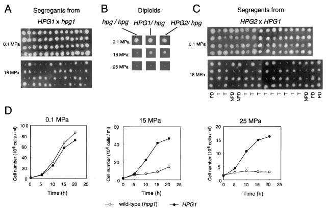
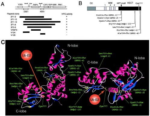
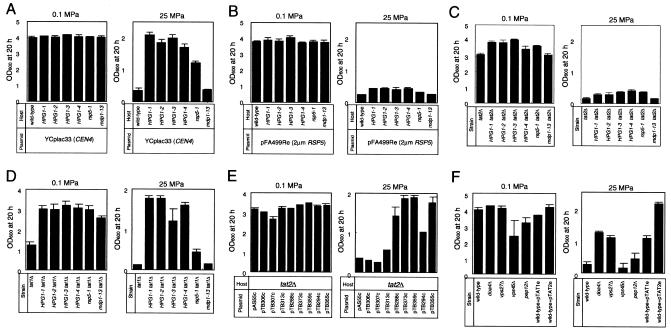
Tryptophan uptake appears to be the Achilles' heel in yeast physiology, since under a variety of seemingly diverse toxic conditions, it becomes the limiting factor for cell growth. When growing cells of Saccharomyces cerevisiae are subjected to high hydrostatic pressure, tryptophan uptake is down-regulated, leading to cell cycle arrest in the G(1) phase. Here we present evidence that the two tryptophan permeases Tat1 and Tat2 are differentially regulated by Rsp5 ubiquitin ligase in response to high hydrostatic pressure. Analysis of high-pressure growth mutants revealed that the HPG1 gene was allelic to RSP5. The HPG1 mutation or the bul1Delta bul2Delta double mutation caused a marked increase in the steady-state level of Tat2 but not of Tat1, although both permeases were degraded at high pressure in an Rsp5-dependent manner. There were marked differences in subcellular localization. Tat1 localized predominantly in the plasma membrane, whereas Tat2 was abundant in the internal membranes. Moreover, Tat1 was associated with lipid rafts, whereas Tat2 localized in bulk lipids. Surprisingly, Tat2 became associated with lipid rafts upon the occurrence of a ubiquitination defect. These results suggest that ubiquitination is an important determinant of the localization and regulation of these tryptophan permeases. Determination of the activation volume (DeltaV( not equal )) for Tat1- and Tat2-mediated tryptophan uptake (89.3 and 50.8 ml/mol, respectively) revealed that both permeases are highly sensitive to membrane perturbation and that Tat1 rather than Tat2 is likely to undergo a dramatic conformational change during tryptophan import. We suggest that hydrostatic pressure is a unique tool for elucidating the dynamics of integral membrane protein functions as well as for probing lipid microenvironments where they localize.


Figures

FIG. 1.
FIG. 1.
Isolation and classification of semidominant HPG mutants. Typical examples of results of high-pressure growth assays are shown. (A) The HPG phenotype of each tetrad derived from the HPG1/hpg1 heterozygous diploid segregated 2 Hpg+:2 Hpg on YPD agar at 18 MPa. The YPD plates were incubated for 2 days at 0.1 or 18 MPa. Note that capital letters are used for the semidominant mutant allele and lowercase letters are used for the wild-type allele. (B) Heterozygous diploids (HPG1/hpg1 and HPG2/hpg2) were grown on YPD agar at 0.1, 18, and 25 MPa for 2 days. (C) Tetrad distribution of segregants derived from an HPG2/HPG2 diploid strain. Segregants were grown on YPD agar at 0.1 and 18 MPa for 2 days. PD, parental ditype; NPD, nonparental ditype; T, tetra type. (D) Cell growth of the wild-type strain and an HPG1 mutant in liquid YPD medium under different pressure conditions. Immediately after decompression, the cell number was counted under a microscope using a hemocytometer.
FIG. 2.
FIG. 2.
HPG1 mutation sites and predicted tertiary structure of the Rsp5 HECT domain. (A) Map of DNA fragments containing RSP5 from an HPG1 genome library that can confer high-pressure growth. (B) Four HPG1 mutations together with some known rsp5 mutation sites are located in the HECT domain of Rsp5. (C) Predicted 3D structure of the Rsp5 HECT domain, obtained by use of 3D-JIGSAW, version 2.0. Predicted α-helices and β-sheets are shown in red and blue, respectively. The HPG1 mutation and some known rsp5 mutation sites are shown in yellow. The ubiquitin-bound E2 enzyme(s) is assumed to have access to the HECT domain from the direction shown in orange.
FIG. 3.
FIG. 3.
Growth properties of HPG1 mutants and some gene disruptants under high-pressure conditions. (A) Cells of the various mutants harboring the empty vector YCplac33 were grown in SC medium at 0.1 and 25 MPa for 20 h. After decompression, the OD600 was determined. (B) Cells of the various mutants containing the wild-type RSP5 gene in a multicopy plasmid were grown at 0.1 and 25 MPa for 20 h. (C through F) Cells of the following mutants were grown at 0.1 and 25 MPa for 20 h: HPG1 tat2Δ mutants (C), HPG1 tat1Δ mutants (D), the tat2Δ mutant harboring TAT2 plasmids containing N-terminal mutations (E), and the doa4Δ, vps27Δ, vps45Δ, and pep12Δ mutants (F). All data are means and standard deviations from three independent experiments.
FIG. 4.
FIG. 4.
Overexpression and deletion of BUL1 have inverse effects. (A) Overexpression of BUL1 but not BUL2 suppresses high-pressure growth of HPG1 mutants. Cells of the indicated genotype containing YEplac195, pBUL1e, or pBUL2e were grown in SC medium at 0.1 and 25 MPa for 20 h. (B) Deletion of BUL1 confers high-pressure growth, but deletion of BUL2 does not. Cells of the wild-type strain and the bul1Δ, bul2Δ, and bul1Δ bul2Δ mutants were grown in SC medium at 0.1 and 25 MPa for 20 h. (C) Cells of the bul1Δ, bul1Δ tat1Δ, bul1Δ tat2Δ, and bul1Δ gap1Δ mutants were grown in SC medium at 0.1 and 25 MPa for 20 h. All data are means and standard deviations from three independent experiments.
FIG. 5.
FIG. 5.
Tat1 and Tat2 are differentially regulated by the HPG1 or bul1Δ bul2Δ mutation. Cells of the indicated mutants containing a functional 3HA-TAT2 or 3HA-TAT1 on YCplac33 (p3HA-TAT2c or p3HA-TAT1c, respectively) were grown in SC medium, and whole-cell extracts were subjected to Western blot analysis. (A) Steady-state levels of Tat1 and Tat2 at 0.1 MPa. (B) Changes in Tat1 and Tat2 levels after the shift to high-pressure conditions at 25 MPa for 2 or 5 h. Typical data from at least four independent experiments are shown. D, degradation; S, stabilization.
FIG. 6.
FIG. 6.
Rsp5 ubiquitin ligase is involved in the subcellular localization of Tat2 but not of Tat1. (A) Subcellular localization of Tat1 and Tat2 by sucrose density gradient centrifugation in wild-type cells. Whole-cell extracts were prepared from the wild-type strain containing either p3HA-TAT1c or p3HA-TAT2c and were subjected to centrifugation on a sucrose density gradient (10 to 60%). Eleven fractions from the top were collected and subjected to Western blot analysis to detect Tat1 and Tat2 together with the four membrane marker proteins Pma1, Pep12, Vps10, and ALP. (B) Subcellular localization of Tat1 and Tat2 in wild-type or mutant cells analyzed by differential centrifugation. Whole-cell extracts prepared from wild-type, HPG1-1, and bu1Δ bul2Δ cells containing either p3HA-TAT1c or p3HA-TAT2c and from Tat25K→R cells (tat2Δ cells containing pTB355c [2HA-Tat25K→R]) were subjected to differential centrifugation to yield the P13 and P100 fractions. Western blotting was performed to detect Tat1 and Tat2 in these fractions together with marker proteins. Typical data from three independent experiments are shown.
FIG. 7.
FIG. 7.
Visualization of ubiquitinated forms of Tat2 (A) or Tat1 (B) in immunoprecipitated proteins present in the P13 and P100 fractions. Cells of the wild-type and HPG1-1 strains containing either YCplac33 (empty vector), p3HA-TAT1c, or p3HA-TAT2c, or tat2Δ cells containing pTB355c (2HA-Tat25K→R), were transformed with YEp105LEU (containing CUP1 promoter-dependent, c-Myc-tagged ubiquitin). After induction of c-Myc-tagged ubiquitin by addition of 0.1 mM CuSO4, whole-cell extracts were prepared and subjected to differential centrifugation to yield the P13 and P100 fractions. Each fraction was solubilized and subjected to immunoprecipitation using an anti-HA antibody-bound affinity matrix as described in Materials and Methods. The immunoprecipitated Tat2 or Tat1 proteins were analyzed by Western blotting using an anti-HA antibody (left) or an anti-c-Myc antibody (right). (Ub)n-Tat2 or -Tat1, ubiquitinated form of Tat2 or Tat1; −, cells containing YEp105LEU and YEplac195 (empty vector); +, cells containing YEp105LEU and either p3HA-TAT2 or p3HA-TAT1; asterisk, nonspecific band.
FIG. 8.
FIG. 8.
Tat2 associates with lipid rafts in ubiquitination-defective cells, whereas Tat1 associates with lipid rafts in an Rsp5-independent manner. (A) Raft association of Tat1 and Tat2 in P13 membrane fractions. Whole-cell extracts from wild-type, HPG1-1, or bul1Δ bul2Δ cells containing either p3HA-TAT1 or p3HA-TAT2, or from tat2Δ cells containing pTB355c (2HA-Tat25K→R), were prepared and subjected to differential centrifugation to yield the P13 fractions. The P13 fractions were solubilized with ice-cold 1% Triton X-100 and subjected to the membrane flotation assay. Four fractions were collected from the top, and the protein samples were analyzed by Western blotting. (B) Raft association of Tat1 and Tat2 in purified plasma membranes. The P13 fractions were prepared as described above and subjected to centrifugation on a sucrose density gradient to yield purified plasma membranes. The plasma membrane samples were solubilized with ice-cold 1% Triton X-100 and subjected to the membrane flotation assay. Four fractions were collected from the top, and the protein samples were analyzed by Western blotting.
FIG. 9.
FIG. 9.
The growth of the tat2Δ mutant is impaired by high concentrations of aromatic amino acids. Cells of the indicated mutants were grown on either SD medium, SC medium, SD medium plus 100 μg of tyrosine/ml (SD+Tyr100), SD medium plus 100 μg of phenylalanine/ml (SD+Phe100), SD medium plus 100 μg of tyrosine/ml and 100 μg of phenylalanine/ml (SD+Try100+Phe100), or YPD agar for 3 days. Typical data from three independent experiments are shown.
FIG. 10.
FIG. 10.
A model for the differential localization and dynamics of the two tryptophan permeases. Tat1 is associated with lipid rafts, whereas Tat2 is localized in bulk lipids. The large ΔVs for Tat1- and Tat2-mediated tryptophan import are accounted for mainly by volume changes associated with protein conformational changes. The initial volume of Tat1 is smaller than that of Tat2 because Tat1 is localized in the highly ordered lipid microdomain of lipid rafts. V, volume of the permease in the initial state; V, volume of the permease in the activated state; ΔV, activation volume associated with tryptophan import through the permease.

References

    1. Abe, F., and K. Horikoshi. 1998. Analysis of intracellular pH in the yeast Saccharomyces cerevisiae under elevated hydrostatic pressure: a study in baro- (piezo-) physiology. Extremophiles 2:223-228. - PubMed
    1. Abe, F., C. Kato, and K. Horikoshi. 1999. Pressure-regulated metabolism in microorganisms. Trends Microbiol. 7:447-452. - PubMed
    1. Abe, F., and K. Horikoshi. 2000. Tryptophan permease gene TAT2 confers high-pressure growth in Saccharomyces cerevisiae. Mol. Cell. Biol. 20:8093-8102. - PMC - PubMed
    1. Abe, F., and K. Horikoshi. 2001. The biotechnological potential of piezophiles. Trends Biotechnol. 19:102-108. - PubMed
    1. Amerik, A. Y., J. Nowak, S. Swaminathan, and M. Hochstrasser. 2000. The Doa4 deubiquitinating enzyme is functionally linked to the vacuolar protein-sorting and endocytic pathways. Mol. Biol. Cell 11:3365-3380. - PMC - PubMed

MeSH terms

Substances