. 2003 Nov;23(21):7648-57.
doi: 10.1128/MCB.23.21.7648-7657.2003.

Miz1 is required for early embryonic development during gastrulation

Affiliations

Miz1 is required for early embryonic development during gastrulation

Sovana Adhikary et al. Mol Cell Biol. 2003 Nov.

Abstract

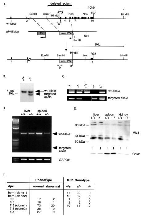
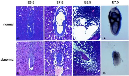
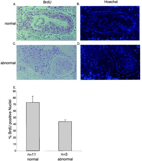
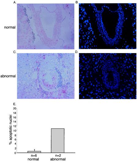
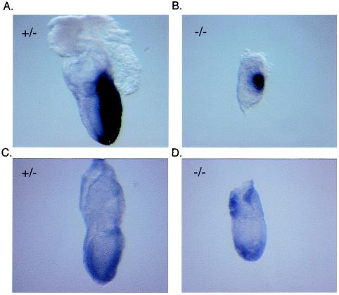
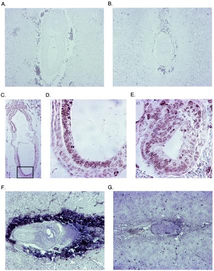
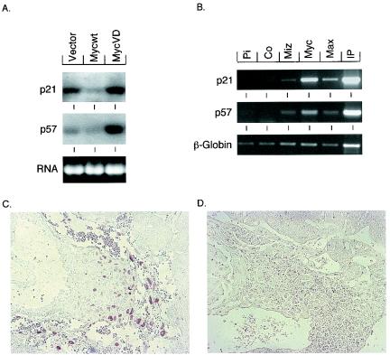
Miz1 is a member of the POZ domain/zinc finger transcription factor family. In vivo, Miz1 forms a complex with the Myc oncoprotein and recruits Myc to core promoter elements. Myc represses transcription through Miz1 binding sites. We now show that the Miz1 gene is ubiquitously expressed during mouse embryogenesis. In order to elucidate the physiological function of Miz1, we have deleted the mouse Miz1 gene by homologous recombination. Miz1(+/-) mice are indistinguishable from wild-type animals; in contrast, Miz1(-/-) embryos are not viable. They are severely retarded in early embryonic development and do not undergo normal gastrulation. Expression of Goosecoid and Brachyury is detectable in Miz1(-/-) embryos, suggesting that Miz1 is not required for signal transduction by Nodal. Expression of p21Cip1, a target gene of Miz1 is unaltered; in contrast, expression of p57Kip2, another target gene of Miz1 is absent in Miz1(-/-) embryos. Miz1(-/-) embryos succumb to massive apoptosis of ectodermal cells around day 7.5 of embryonic development. Our results show that Miz1 is required for early embryonic development during gastrulation.


Figures

FIG. 1.
FIG. 1.
Expression of Miz1 during embryogenesis. (A and B) Whole-mount in situ hybridization documenting expression of Miz1 mRNA in embryos at E6.5 (A) and E9.5 (B). In each panel, the embryo on the left was hybridized with an antisense probe, and the embryo on the right was hybridized with a sense control probe. (C) In situ hybridization documenting expression of Miz1 mRNA in a section of an E13.5 embryo. The inserts show hybridization with a sense probe as the negative control, on the left side illuminated as the antisense section and on the right side as a dark-field picture. (D and E) In situ hybridization documenting expression of Miz1 mRNA in epithelia of an E15.5 embryo. The pictures show a sagittal section of the nose with primordia of the vibrissae (D) and a sagittal section of the tongue (E).
FIG. 2.
FIG. 2.
Disruption of the Miz1 gene. (A) Targeting strategy. The panel shows the structure of the murine genomic locus of Miz1, the replacement vector (pPNT-Miz1), and the structure of the targeted allele. (B) Southern blot documenting successful targeting of the Miz1 locus in ES cells. Shown is a hybridization using the probe indicated in panel A to a HindIII digest of genomic DNA. The expected fragments are shown in panel A. (C) PCR-based genotyping of E7.5 embryos using primer pairs specific for either the wild-type (wt) or the targeted Miz1 locus. The primers used are indicated in panel A as “a” for the wild-type and “b” for the knockout allele. Shown are results derived from intercrosses of Miz1+/− animals. (D) Absence of a transcript from the targeted allele. Shown are results from reverse transcriptase PCR assays using primers located in exons 2 and 7 of Miz1 from RNA isolated from liver and spleen of heterozygous animals. An arrow indicates the predicted size of the PCR product from the knockout allele. (E) Absence of a truncated protein in heterozygous embryos. The panel shows a Western blot of lysates of the indicated organs from either wild-type or Miz1+/− animals with an anti-Miz1 monoclonal antibody (10E2). (F) Phenotypes and genotype of neonates and embryos from the heterozygous intercrosses. Many of the abnormal embryos could not be genotyped due to their size and/or beginning resorption. Therefore, the ratios of the genotypes are skewed. dpc, day postcoitus.
FIG. 3.
FIG. 3.
Phenotype of Miz1−/− embryos. Histological sections of embryos generated from crosses between Miz1 heterozygous mice. (A and B) Sagittal section of E6.5 normal (A) and abnormal (B) embryos. (C and D) Sagittal section of E7.5 normal (C) and abnormal (D) embryos. (E and F) Transverse section of E8.5 embryo, which had completed the process of turning (E), and sagittal section of E8.5 mutant embryo in the process of resorption (F). The abnormal embryo (F) is depicted in a twofold-higher magnification than its normal littermate. (G and H) Whole-mount RNA in situ hybridization of normal and mutant embryos with a Miz1 probe.
FIG. 4.
FIG. 4.
BrdU incorporation in E7.0 embryos. Panels A and C document staining of sections of normal (A) and abnormal (C) E7.5 embryos with antibodies directed against BrdU; animals were injected with BrdU 2 h before fixation. The right panels (B and D) show counterstaining with Hoechst 33285 to visualize nuclei. Panel E shows a quantitation of the results obtained by calculating the percentage of BrdU-incorporating cells of 11 normal and 3 abnormal embryos. The error bars represent the standard deviation.
FIG. 5.
FIG. 5.
Apoptosis in E7.5 embryos. Panels A and C document TUNEL staining of morphologically normal (A) and abnormal (C) embryos at E7.5. Panels B and D document staining of the nuclei with Hoechst 33285. Panel E shows a quantitation of the results. The percentages of apoptotic nuclei were calculated for six normal and two abnormal embryos.
FIG. 6.
FIG. 6.
Expression of Brachyury and Goosecoid in E7.5 embryos. The panels document whole-mount in situ hybridization with either a Brachyury (A and B) or a Goosecoid (C and D) antisense probe of E7.5 embryos genotyped as Miz1−/− (B and D) and of two of their Miz1+/− littermates (A and C).
FIG. 7.
FIG. 7.
Expression of p15Ink4b, p21Cip1, and c-myc in normal and abnormal mouse embryos. The upper two panels document staining with an anti-p15Ink4b antibody of either a normal (A) or an abnormal (B) antibody. Note the absence of staining in the embryo, but the presence of staining in the surrounding decidua. The middle panels show staining of morphologically normal (C and D) or abnormal (E) E7.5 embryos with an anti-p21Cip1 antibody. The embryo in panel C is shown in a lower magnification; the frame marks the area shown in panel D at the same magnification as the abnormal embryo in section E. The lower two panels show in situ hybridizations with a c-myc antisense probe of either a normal (F) or an abnormal (G) E7.5 embryo. Note that the intense staining surrounding the normal embryo derives from the peroxidase present in erythrocytes and does not reflect c-myc expression.
FIG. 8.
FIG. 8.
p57Kip2 is a target gene of Miz1 and is absent in Miz−/− embryos. (A) Northern blots demonstrating differential expression of the indicated genes in primary mouse embryo fibroblasts infected with viruses of the indicated genotypes. MycV394D is an allele of Myc that does not bind to Miz1 (20). (B) ChIp experiments documenting in vivo binding of Myc and Miz1 to DNA surrounding the start site of transcription of the P57KIP2and P21CIP1 genes. For P57KIP2, a fragment between nucleotides −139 and +65 relative to the major start site of transcription was amplified, and for P21CIP1, a fragment between nucleotides −61 and +353 was amplified. The experiment was performed with HeLa cells as described by Herold et al. (20). Panels C and D document staining of the ectoplacental cone of morphologically normal (C) or abnormal (D) E7.5 embryos with anti-p57Kip2 antibodies.

Similar articles

Cited by

References

    1. Amati, B., M. W. Brooks, N. Levy, T. D. Littlewood, G. I. Evan, and H. Land. 1993. Oncogenic activity of the c-Myc protein requires dimerization with Max. Cell 72:233-245. - PubMed
    1. Arnold, I., and F. M. Watt. 2001. c-Myc activation in transgenic mouse epidermis results in mobilization of stem cells and differentiation of their progeny. Curr. Biol. 11:558-568. - PubMed
    1. Bancroft, J. D., and A. Stevens. 1990. Theory and practice of histological techniques, 3rd ed. Churchill Livingstone, Edinburgh, United Kingdom.
    1. Bardwell, V. J., and R. Treisman. 1994. The POZ domain: a conserved protein-protein interaction motif. Genes Dev. 8:1664-1677. - PubMed
    1. Belo, J. A., T. Bouwmeester, L. Leyns, N. Kertesz, M. Gallo, M. Follettie, and E. M. De Robertis. 1997. Cerberus-like is a secreted factor with neutralizing activity expressed in the anterior primitive endoderm of the mouse gastrula. Mech. Dev. 68:45-57. - PubMed

Publication types

MeSH terms

Substances