. 2003 Oct;24(9):1757-64.

Earring lesions of the parotid tail

Affiliations

Earring lesions of the parotid tail

Bronwyn E Hamilton et al. AJNR Am J Neuroradiol. 2003 Oct.

Abstract

Background and purpose: Masses in the parotid tail can be a source of consternation to radiologists and clinicians; inaccurate localization may lead to significant iatrogenic complication. We sought to review the pertinent anatomic localizing features of the parotid tail, relevant facial nerve anatomy, and sources of clinical and radiologic confusion. To conclude, we review imaging features that are helpful in generating a diagnosis in this location.

Methods: We retrospectively reviewed the imaging and clinical features of 111 parotid tail masses in 103 patients (56 male, 45 female, two of unknown sex; age range, 5 months-81 years). The following imaging findings were noted: size, enhancement, multiplicity of lesions, attenuation on CT scans, signal intensity on MR images, and appearance of the surrounding parotid gland. Diagnosis was confirmed by either surgical resection or biopsy findings or by specific clinical data or characteristic imaging findings.

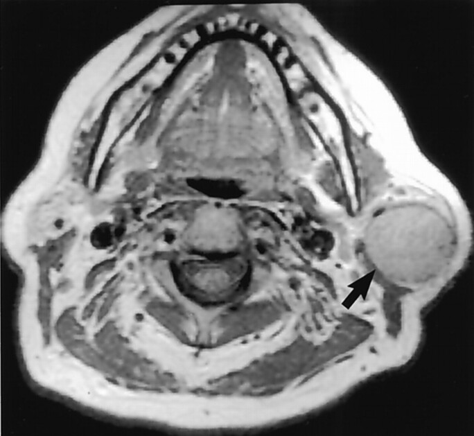
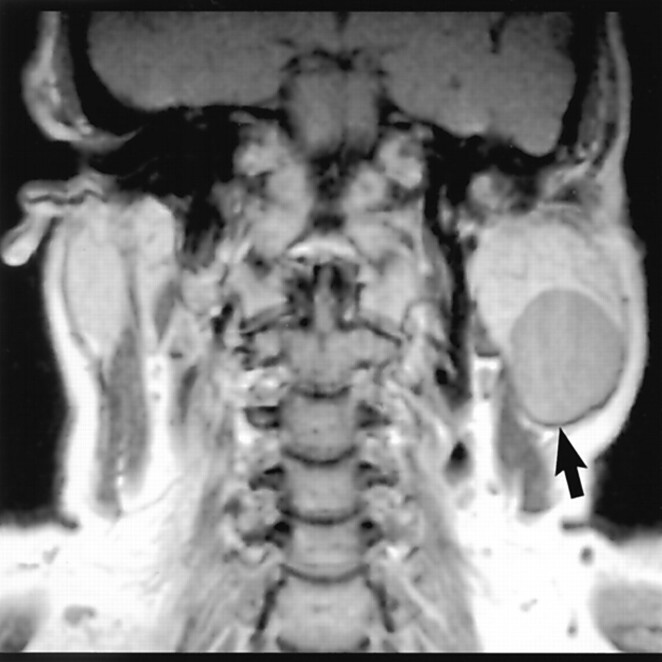
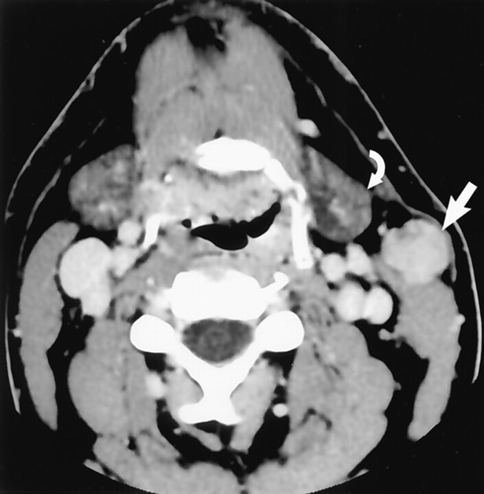
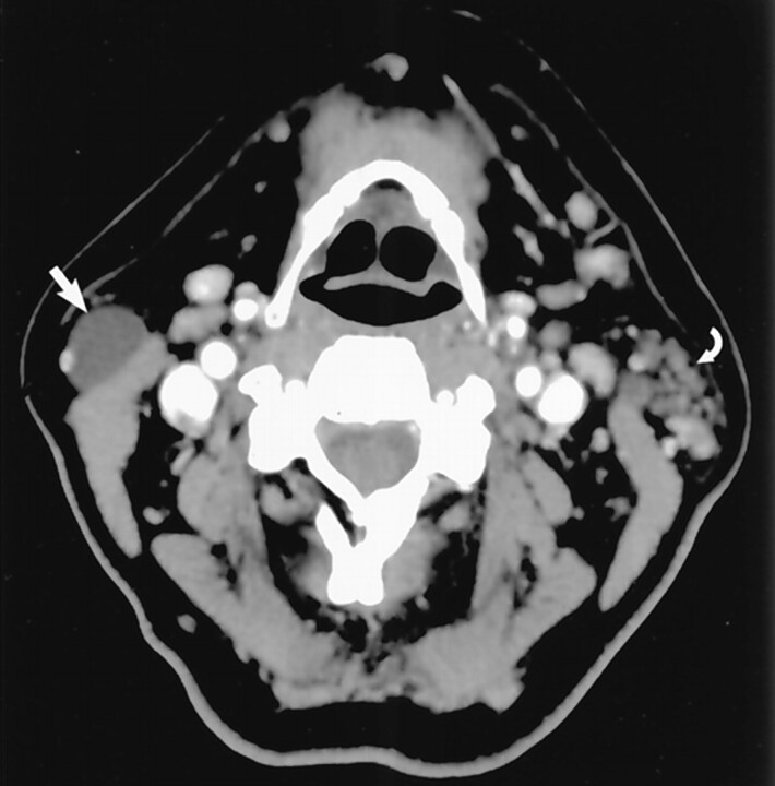
Results: Seventeen types of parotid tail masses were identified. Benign lesions were: pleomorphic adenoma (n = 15), Warthin tumor (n = 14), infectious process (n = 13), venous malformation (n = 9), and Sjögren disease (n = 9), lymphatic malformations (n = 7), lipoma (n = 6), HIV lymphoepithelial lesion (n = 4), first brachial cleft cyst (n = 3), oncocytoma (n = 2), sarcoid (n = 1), and lymph node (n = 1). Malignant lesions were: Non-Hodgkin lymphoma (n = 14), metastatic disease (n = 7), mucoepidermoid carcinoma (n = 4), acinic cell carcinoma (n = 1), and undifferentiated carcinoma (n = 1). Eight patients had two diagnoses.

Conclusion: Understanding normal parotid tail anatomy is important to radiologists, because accurate localization has implications for appropriate management of masses in this location, potentially reducing the occurrence of marginal mandibular nerve injury.


Figures

F<sc>ig</sc> 1.
Fig 1.
Diagram of parotid gland with a typical mass in the tail. Course of the facial nerve is demonstrated. Note the relationship of the marginal mandibular nerve (arrow) as it traverses the parotid tail mass. (Reprinted with permission from reference .)
F<sc>ig</sc> 2.
Fig 2.
Axial contrast-enhanced CT scan demonstrates normal parotid tail anatomy. Anatomic boundaries of the parotid tail are the following: lateral and superficial boundary, platysma (white arrow); anteromedial boundary, posterior belly of digastric (black arrow); posteromedial boundary, SCM (curved white arrow). (Reprinted with permission from reference .)
F<sc>ig</sc> 3.
Fig 3.
Axial contrast-enhanced CT scan demonstrates normal jugulodigastric lymph nodes bilaterally (curved arrows), which should not be confused with lesions originating in the parotid tail. Note the normal parotid tail tissue located anterolateral to the SCM (straight arrow). The lymph nodes lie anteromedial to the muscle. (Reprinted with permission from reference .)
F<sc>ig</sc> 4.
Fig 4.
Earring lesion of the parotid tail. Axial contrast-enhanced CT scan shows typical appearance of a cystic Warthin tumor. Note the nodule of soft tissue (straight white arrow), which is commonly seen. A small amount of parotid tissue (open arrow) is seen anterior to the mass. Note parotid tail anatomic landmarks: SCM posteriorly and platysma (curved white arrow) laterally. (Reprinted with permission from reference .)
F<sc>ig</sc> 5.
Fig 5.
Axial nonenhanced T1-weighted MR image demonstrates a large mass (arrow) in the left parotid tail. This mass has little inherent contrast against adjacent normal tissues, making localization more challenging. (Reprinted with permission from reference .)
F<sc>ig</sc> 6.
Fig 6.
Coronal nonenhanced T1-weighted MR image clarifies the intraparotid nature of this BMT (arrow). (Reprinted with permission from reference .)
F<sc>ig</sc> 7.
Fig 7.
Mass was believed to represent a submandibular space or gland tumor by the referring general surgeon, who initially planned to excise the mass. Axial contrast-enhanced CT image reveals the mass (straight arrow) to be deep to platysma and anterior to the SCM, characteristic for a parotid tail “earring lesion.” Note its proximity to the submandibular gland (curved arrow). (Reprinted with permission from reference .)
F<sc>ig</sc> 8.
Fig 8.
Otolaryngology referred this patient for imaging after palpating a mass in the “left parotid tail.” Axial contrast-enhanced CT scan through the mass reveals a partly cystic mass in a typical location for a jugulodigastric lymph node (curved arrow). Normal parotid tail tissue is seen (straight white arrow). This was a metastatic node from a clinically occult primary squamous cell carcinoma, which is partly seen on this section in the left faucial tonsil (open arrow). Additional lymph nodes lower in the neck indicated a high-stage tumor. Upon chart review, the referring physician had noted mass effect in the left tonsil; however, a mucosal lesion was not present. (Reprinted with permission from reference .)
F<sc>ig</sc> 9.
Fig 9.
Axial contrast-enhanced CT scan through the parotid tail demonstrates a dominant cyst (straight arrow). Nodular lymphoid aggregates (curved arrow) in the contralateral gland should suggest a systemic process, in this case, Sjögren disease. The appearance is typical of Sjögren disease or HIV lymphoepithelial lesion, which cannot be distinguished on a histopathologic basis. (Reprinted with permission from reference .)

References

    1. Harnsberger HR. Head and Neck Digital Teaching File. Salt Lake City, UT: Amirsys, Inc.;2002
    1. Bannister LH. Alimentary system. In: Williams PL, Bannister L, Berry M, et al, eds. Gray’s Anatomy: The Anatomical Basis of Medicine and Surgery. 38th ed. Edinburgh, Scotland: Churchill Livingstone1995. :1691–1692
    1. Som PM, Brandwein M. Salivary glands. In: Som PM, Curtin HD, eds. Head and Neck Imaging. 3rd ed, vol 2. St. Louis, MO: Mosby-Year Book, Inc.;1996. :824–830, 898–907
    1. Hoffman H, Funk G, Endres D. Evaluation and surgical treatment of tumors of the salivary glands. In: Thawley SE, Panje W, Batsakis L, Robert J, eds. Comprehensive Management of Head and Neck Tumors. 2nd ed, vol 2. Philadelphia, PA: W. B. Saunders Company;1999. :1147–1177
    1. Gates G, Johns M. Embryology and anatomy of the salivary glands. In: Paparella MM, Shumrick D, eds. Otolaryngology: Basic Sciences and Related Disciplines. 2nd ed, vol 1. Philadelphia, PA: W. B. Saunders Company;1980. :150–151

MeSH terms

LinkOut - more resources