Case Reports
. 2003 Oct;24(9):1789-96.

Endovascular treatment strategy for direct carotid-cavernous fistulas resulting from rupture of intracavernous carotid aneurysms

Affiliations
Case Reports

Endovascular treatment strategy for direct carotid-cavernous fistulas resulting from rupture of intracavernous carotid aneurysms

Nozomu Kobayashi et al. AJNR Am J Neuroradiol. 2003 Oct.

Abstract

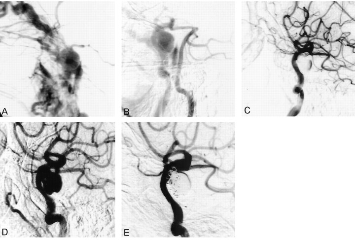
Background and purpose: Reported treatments and outcomes in aneurysmal carotid-cavernous fistulas (CCFs) have been admixed with those of cases considered to be symptomatic of intracavernous aneurysm. However, aneurysmal CCFs have clinical features distinct from those of dural arteriovenous fistulas, and treatment strategies similar to those of traumatic CCF are required. We evaluated our experience in placing detachable balloons in the management of spontaneous CCFs due to rupture of an intracavernous aneurysm.

Methods: Six patients (one man, five women; mean age, 64.7 years) were treated for spontaneous direct CCF at our institution between 1995 and 2001. All patients presented with sudden ocular symptoms including exophthalmos, conjunctival injection, chemosis, and ocular motor palsies. Detachable latex balloons were used as the embolic material in five patients, and in one patient the cavernous sinus was packed transarterially with coils.

Results: All six patients were successfully treated by means of transarterial embolization, and symptoms improved within a week.

Conclusion: Although other techniques using a transvenous approach and/or detachable coils may also be useful, embolization with detachable balloons should be a safe and effective method to immediately occlude the fistula.


Figures

F<sc>ig</sc> 1.
Fig 1.
Case 1. A, MR angiogram reveals an aneurysm (arrow) involving the right ICA. B–D, Angiograms show a right direct CCF with high-flow shunt (B), which clinically was symptomatic. The aneurysm is first opacified in the early phase (C). Obliteration of the shunt is achieved (D). E, Angiogram obtained 2 years later reveals partial recurrence of the aneurysm
F<sc>ig</sc> 2.
Fig 2.
Case 2. A, Carotid angiogram shows a direct CCF; the distal portion of the ICA is not opacified because of the high-flow shunt. B, Right vertebral angiogram depicts the aneurysmal sac by collateral flow upon cross compression. C, Carotid angiogram obtained just after the procedure shows that a detachable balloon completely occludes the shunt. D and E, Angiograms obtained 3 months later reveal recurrence of the aneurysm (D), which is packed with coils (E)
F<sc>ig</sc> 3.
Fig 3.
Case 3. A and B, Left carotid angiograms show a high-flow CCF (A). Shunt flow is nearly obliterated by a detachable balloon, but the orifice is too large to permit complete obliteration. In addition, the space between the orifice and the wall of cavernous sinus is too small to safely position a sufficiently inflated balloon at the fistula (B); further inflation of the balloon can result in migration into the parent artery. Therefore, parent artery occlusion was performed after confirming cross flow with a test occlusion.
F<sc>ig</sc> 4.
Fig 4.
Case 4. A and B, Left carotid angiograms show a high-flow CCF (A), which is obliterated by a detachable balloon (B). C and D, Follow-up angiograms obtained 1 month later reveal recurrence of the aneurysmal sac (C), which was embolized with detachable coils (D).
F<sc>ig</sc> 5.
Fig 5.
Case 5. A, Left carotid angiogram shows a direct CCF. B and C, Oblique views show that the small aneurysm is opacified initially, and then shunt flow is seen. D–F, Delivery of the detachable balloon fails because of a small orifice, and a microcatheter is advanced into the cavernous sinus (D). The sinus is packed with coils (E), producing nearly complete obliteration (F).
F<sc>ig</sc> 6.
Fig 6.
Case 6. A, Right carotid angiogram shows a direct CCF mainly draining to the superior ophthalmic vein. A microcatheter is inserted into the ruptured aneurysmal sac. B, Fistula disappears after treatment with a detachable balloon, without impairing carotid artery flow.

References

    1. Barrow DL, Spector RH, Braun IF, Landman JA, Tindall SC, Tindall GT. Classification and treatment of spontaneous carotid-cavernous sinus fistulas. J Neurosurg 1985;62:248–256 - PubMed
    1. Halbach VV, Higashida RT, Hieshima GB, Hardin CW, Yang PJ. Transvenous embolization of direct carotid cavernous fistulas. AJNR Am J Neuroradiol 1988;9:741–747 - PMC - PubMed
    1. Kanner AA, Maimon S, Rappaport ZH. Treatment of spontaneous carotid-cavernous fistula in Ehlers-Danlos syndrome by transvenous occlusion with Guglielmi detachable coils: case report. J Neurosurg 2000;93:689–692 - PubMed
    1. Debrun GM, Aletich VA, Miller NR, DeKeiser RJW. Three cases of spontaneous direct carotid cavernous fistulas associated with Ehlers-Danlos syndrome type IV. Surg Neurol 1996;46:247–252 - PubMed
    1. d’Angelo VA, Monte V, Scialfa G, Fiumara E, Scotti G. Intracerebral venous hemorrhage in “high-risk” carotid-cavernous fistula. Surg Neurol 1988;30:387–390 - PubMed

Publication types

MeSH terms