

Intracranial aneurysms treated with Guglielmi detachable coils: is contrast material necessary in the follow-up with 3D time-of-flight MR angiography?
- PMID: 14561605
- PMCID: PMC7976302
Intracranial aneurysms treated with Guglielmi detachable coils: is contrast material necessary in the follow-up with 3D time-of-flight MR angiography?
Abstract
Background and purpose: Three-dimensional time-of-flight (TOF) MR angiography has been evaluated in the follow-up of intracranial aneurysms treated with Guglielmi detachable coils (GDCs) with good results. Some of the studies used contrast material in addition to the 3D TOF MR technique and others did not. We assessed the usefulness of contrast material with 3D TOF MR angiography by comparing this sequence before and after contrast material injection.
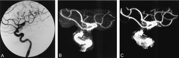
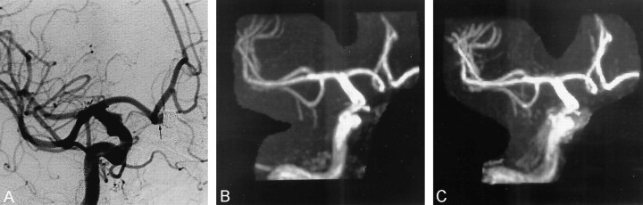
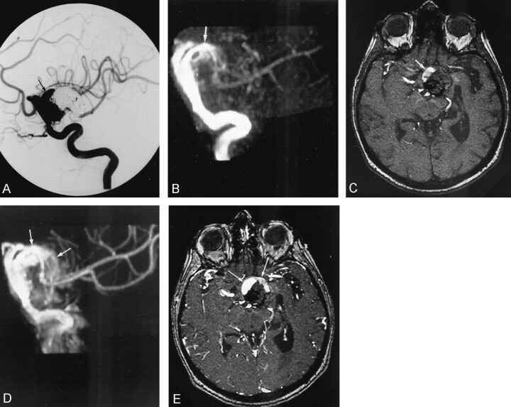
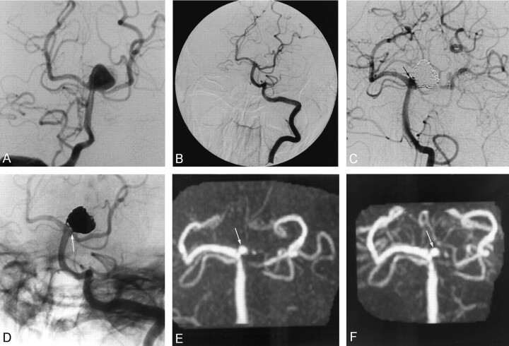
Methods: Fifty-eight patients harboring a total of 71 cerebral aneurysms previously treated with GDCs were included in the prospective study. MR angiography (at 1.5 T) was performed with a 3D TOF sequence before and after injection of gadolinium-based contrast material. Features evaluated were presence and size of a neck remnant, parent and adjacent vessel patency, and venous overlap. Digital subtraction angiography was the standard of reference.
Results: Comparison of the techniques showed a good agreement in the detection of residual flow. Six cases of small residual neck were not detected with either the 3D TOF or the contrast-enhanced 3D TOF sequence. In one case of giant aneurysm, the extent of recanalization was more evident after contrast material administration. The use of contrast material did not help to show the parent and adjacent arteries. Venous overlap on contrast-enhanced 3D TOF angiograms did not affect image interpretation.
Conclusion: In this series, the use of intravenous contrast material did not improve the ability of 3D TOF MR angiography to depict the presence of residual or recurrent aneurysms previously treated with endovascular coiling. In one giant aneurysm, use of intravenous contrast material did result in improved visualization of a residual aneurysm.
Figures
References
-
- Vinuela F, Duckwiler G, Mawad M, Guglielmi G. Detachable coil embolization of acute intracranial aneurysm: perioperative anatomical and clinical outcome in 403 patients. J Neurosurg 1997;86:475–482 - PubMed
-
- Kähära VJ, Seppänen SK, Ryymin PS, Mattila P, Kuurne T, Laasonen EM. MR angiography with three-dimensional time-of-flight and targeted maximum-intensity-projection reconstructions in the follow-up of intracranial aneurysms embolized with Guglielmi detachable coils. AJNR Am J Neuroradiol 1999;20:1470–1475 - PMC - PubMed
-
- Brunereau L, Cottier JP, Sonier CB, et al. Prospective evaluation of time-of-flight MR angiography in the follow-up of intracranial saccular aneurysms treated with Guglielmi detachable coils. J Comput Assist Tomogr 1999;23:216–223 - PubMed
MeSH terms
Substances
LinkOut - more resources
Full Text Sources
Medical