

Relationship between the concentration of supplemental oxygen and signal intensity of CSF depicted by fluid-attenuated inversion recovery imaging
- PMID: 14561617
- PMCID: PMC7976282
Relationship between the concentration of supplemental oxygen and signal intensity of CSF depicted by fluid-attenuated inversion recovery imaging
Abstract
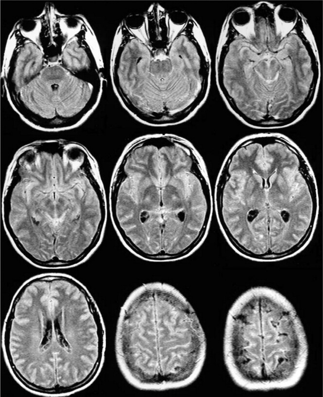
Background and purpose: Prior reports have described increased signal intensity (SI) of CSF on fluid-attenuated inversion recovery (FLAIR) images of anesthetized patients receiving 100% O(2). This appearance can simulate that of diseases. We evaluated the relationship between the concentration of inhaled O(2) and the development of increased SI of CSF on FLAIR images.
Methods: FLAIR was performed in 25 healthy volunteers breathing room air and 100% O(2) through a face mask for 5, 10, and 15 minutes. MR imaging, including FLAIR imaging, was performed in 52 patients with no potential meningeal abnormalities under general anesthesia: 21 received an equal mixture of N(2)O and O(2), and 31 received 100% O(2). The SI of CSF in volunteers and patients was graded in several locations by using a three-point scale.
Results: SI of CSF significantly increased (P <.05) in various locations, in both volunteers and patients breathing 100% O(2), when compared with SI in the same volunteers breathing room air. Hyperintensity of CSF was not significantly different in volunteers receiving 100% O(2) through a face mask compared with anesthetized patients receiving 100% O(2) through a laryngeal airway or an endotracheal tube. No significant increase in SI occurred in patients receiving 50% O(2), when compared with the SI of volunteers breathing room air.
Conclusion: Supplemental oxygen at 100% is a main cause of artifactual CSF hyperintensity on FLAIR images, regardless of the anesthetic drug used. This artifact does not develop when 50% O(2) is administered.
Figures


Similar articles
-
Supplemental oxygen causes increased signal intensity in subarachnoid cerebrospinal fluid on brain FLAIR MR images obtained in children during general anesthesia.Radiology. 2004 Oct;233(1):51-5. doi: 10.1148/radiol.2331031375. Radiology. 2004. PMID: 15454616 Clinical Trial.
-
Hyperintense signal abnormality in subarachnoid spaces and basal cisterns on MR images of children anesthetized with propofol: new fluid-attenuated inversion recovery finding.AJNR Am J Neuroradiol. 2001 Feb;22(2):394-9. AJNR Am J Neuroradiol. 2001. PMID: 11156789 Free PMC article.
-
Fraction of inspired oxygen in relation to cerebrospinal fluid hyperintensity on FLAIR MR imaging of the brain in children and young adults undergoing anesthesia.AJR Am J Roentgenol. 2002 Sep;179(3):791-6. doi: 10.2214/ajr.179.3.1790791. AJR Am J Roentgenol. 2002. PMID: 12185066
-
Abnormal hyperintensity within the subarachnoid space evaluated by fluid-attenuated inversion-recovery MR imaging: a spectrum of central nervous system diseases.Eur Radiol. 2003 Dec;13 Suppl 4:L192-201. doi: 10.1007/s00330-003-1877-9. Eur Radiol. 2003. PMID: 15018187 Review.
-
Fluid-attenuated inversion recovery (FLAIR): clinical prospectus of current and future applications.Top Magn Reson Imaging. 1996 Dec;8(6):389-96. Top Magn Reson Imaging. 1996. PMID: 9402679 Review.
Cited by
-
Carotid stent delivery in an XMR suite: immediate assessment of the physiologic impact of extracranial revascularization.AJNR Am J Neuroradiol. 2005 Mar;26(3):531-7. AJNR Am J Neuroradiol. 2005. PMID: 15760861 Free PMC article.
-
The effects of normobaric and hyperbaric oxygenation on MRI signal intensities in T1 -weighted, T2 -weighted and FLAIR images in human brain.Radiol Oncol. 2023 Sep 4;57(3):317-324. doi: 10.2478/raon-2023-0043. eCollection 2023 Sep 1. Radiol Oncol. 2023. PMID: 37665738 Free PMC article.
-
Reduction of Oxygen-Induced CSF Hyperintensity on FLAIR MR Images in Sedated Children: Usefulness of Magnetization-Prepared FLAIR Imaging.AJNR Am J Neuroradiol. 2016 Aug;37(8):1549-55. doi: 10.3174/ajnr.A4723. Epub 2016 Mar 17. AJNR Am J Neuroradiol. 2016. PMID: 26988816 Free PMC article.
-
Noninvasive MR cisternography with fluid-attenuated inversion recovery and 100% supplemental O(2) in the evaluation of neurocysticercosis.AJNR Am J Neuroradiol. 2004 Feb;25(2):295-7. AJNR Am J Neuroradiol. 2004. PMID: 14970035 Free PMC article.
-
Dynamic oxygen-enhanced MRI of cerebrospinal fluid.PLoS One. 2014 Jun 23;9(6):e100723. doi: 10.1371/journal.pone.0100723. eCollection 2014. PLoS One. 2014. PMID: 24956198 Free PMC article.
References
-
- Deliganis AV, Fisher DJ, Lam AM, Maravilla KR. Cerebrospinal fluid signal intensity increase on FLAIR MR images in patients under general anesthesia: the role of supplemental O2. Radiology 2001;218:152–156 - PubMed
-
- Weinberger E, Frigon C, Astley S, Jardine D, Shaw DW. Subarachnoid CSF Hyperintensity on Brain MR FLAIR images in anesthetized patients. Pediatric Radiol 2001;31:S1
-
- Dechambre SD, Duprez T, Grandin CB, Lecouvet FE, Peeters A, Cosnard G. High signal in cerebrospinal fluid mimicking subarachnoid haemorrhage on FLAIR following acute stroke and intravenous contrast medium. Neuroradiology 2000;42:608–611 - PubMed
-
- Noguchi K, Ogawa T, Seto H, et al. Subacute and chronic subarachnoid hemorrhage: diagnosis with fluid-attenuated inversion-recovery MR imaging. Radiology 1997;203:257–262 - PubMed
MeSH terms
Substances
LinkOut - more resources
Full Text Sources
Other Literature Sources
Medical