

MR imaging characteristics of pilomyxoid astrocytomas
- PMID: 14561626
- PMCID: PMC7976309
MR imaging characteristics of pilomyxoid astrocytomas
Abstract
Background and purpose: Pilomyxoid astrocytoma (PMA) is a recently described tumor that typically occurs in the chiasmatic-hypothalamic region in young children and has unique histopathologic and clinical characteristics. These tumors have been previously diagnosed as pilocytic astrocytoma (PA). PMA appears to have a higher rate of recurrence and CSF dissemination than typical PA.
Methods: We analyzed MR findings in four patients with PMA and compared them with those of typical chiasmatic-hypothalamic PA.
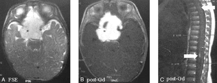
Results: MR findings of PMA were chiasmatic or hypothalamic enhancing solid tumor with hydrocephalus, highly homogeneous T2 signal intensity that extended into the deep white and gray matter, and CSF dissemination.
Conclusion: Larger series are needed before the MR imaging findings of chiasmatic or hypothalamic enhancing solid tumor with hydrocephalus, highly homogeneous T2 signal intensity extending into the deep white and gray matter, and CSF dissemination can be used in the differential diagnosis of such tumors.
Figures
References
-
- Tihan T, Fisher PG, Kepner JL, et al. Pediatric astrocytomas with monomorphous pilomyxoid features and a less favorable outcome. J Neuropathol Exp Neurol 1999;58:1061–1068 - PubMed
-
- Fuller CE, Frankel B, Smith M, et al. Suprasellar monomorphous pilomyxoid neoplasm: an ultrastructural analysis. Clin Neuropathol 2001;20:256–262 - PubMed
-
- Burger P. Pathology of brain stem astrocytomas. Pediatr Neurosurg 1996;24:35–40 - PubMed
-
- Krieger MD, Gonzales-Gomez I, Levy ML, McComb JG. Recurrence patterns and anaplastic change in a long term study of pilocytic astrocytomas. Pediatr Neurosurg 1997;27:1–11 - PubMed
-
- Burger PC, Cohen KJ, Rosenblum MK, Tihan T. Pathology of diencephalic astrocytomas. Pediatr Neurosurg 2000;32:214–219 - PubMed
MeSH terms
LinkOut - more resources
Full Text Sources
Medical