Case Reports
. 2003 Oct;24(9):1923-5; author reply 1925.

Prenatal diagnosis of a vein of Galen aneurysmal malformation with fetal MR imaging study

Case Reports

Prenatal diagnosis of a vein of Galen aneurysmal malformation with fetal MR imaging study

Anna Messori et al. AJNR Am J Neuroradiol. 2003 Oct.
No abstract available


Figures

F<sc>ig</sc> 1.
Fig 1.
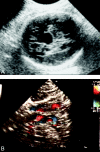
Ultrasonogram and color Doppler sonogram from the case of a 34-year-old gravida 2, para 0 woman who was referred at 38 weeks and 5 days of gestation. A, Routine ultrasonogram shows an intracranial echo-free area. B, Color Doppler sonogram shows turbulent blood flow within the lesion (courtesy of Dr. G. Ciotti, Department of Obstetrics, Children’s Hospital “G. Salesi,” Ancona, Italy).
F<sc>ig</sc> 2.
Fig 2.
Fetal MR images obtained at 38 weeks and 5 days of gestation (body coil; fast low angle shot 2D; 44/10 [TR/TE]; flip angle, 40 degrees; section thickness, 5 mm; field of view, 380 mm; matrix, 192 × 256). A, Midsagittal view of the fetal brain shows a distended midline vascular structure; the brain parenchyma appears otherwise normal, and there is no ventricular dilation. B, Axial view.
F<sc>ig</sc> 3.
Fig 3.
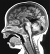
Postnatal midsagittal T1-weighted spin-echo MR image obtained at 75 days of life (500/15; section thickness, 5 mm; field of view, 230 mm; matrix, 220 × 256) shows an interhemispheric vascular malformation at the level of the posterior third and splenium of the corpus callosum, flowing into a markedly distended vein of Galen and into the straight sinus.
F<sc>ig</sc> 4.
Fig 4.
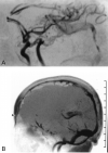
Postnatal MR angiograms obtained at 75 days of life. A, Arterial flow (fast imaging with steady-state free precession 3D, magnetization transfer-tilted optimized nonsaturating excitation, 44/10). B, Venous flow (fast low angle shot 2D, 40/10). Although turbulent flow through the vein of Galen makes it impossible to directly show it, the images indirectly show the malformation via its arterial supply and the venous flow into the straight sinus.

Comment on

References

    1. Jones BV, Ball WS, Tomsick TA, Millard J, Crone KR. Vein of Galen aneurysmal malformation: diagnosis and treatment of 13 children with extended clinical follow-up. AJNR Am J Neuroradiol 2002;23:1717–1724 - PMC - PubMed
    1. Yuh WT, Nguyen HD, Fisher DJ, et al. MR of fetal central nervous system abnormalities. AJNR Am J Neuroradiol 1994;15:459–464 - PMC - PubMed
    1. Yamashita Y, Abe T, Ohara N, et al. Successful treatment of neonatal aneurysmal dilatation of the vein of Galen: the role of prenatal diagnosis and trans-arterial embolization. Neuroradiology 1992;34:457–459 - PubMed
    1. Campi A, Scotti G, Filippi M, Gerevini S, Strigimi F, Lasjaunias P. Antenatal diagnosis of vein of Galen aneurysmal malformation: MR study of fetal brain and postnatal follow-up. Neuroradiology 1996;38:87–90 - PubMed
    1. Kurihara N, Tokieda K, Ikeda K, et al. Prenatal MR findings in a case of aneurysm of the vein of Galen. Pediatr Radiol 2001;31:160–162 - PubMed

MeSH terms