. 2003 Oct;112(8):1142-51.
doi: 10.1172/JCI18549.

Endothelial and nonendothelial sources of PDGF-B regulate pericyte recruitment and influence vascular pattern formation in tumors

Affiliations

Endothelial and nonendothelial sources of PDGF-B regulate pericyte recruitment and influence vascular pattern formation in tumors

Alexandra Abramsson et al. J Clin Invest. 2003 Oct.

Abstract

Tumor-infiltrating blood vessels deviate morphologically and biochemically from normal vessels, raising the prospect of selective pharmacological targeting. Current antiangiogenic approaches focus mainly on endothelial cells, but recent data imply that targeting pericytes may provide additional benefits. Further development of these concepts will require deeper insight into mechanisms of pericyte recruitment and function in tumors. Here, we applied genetic tools to decipher the function of PDGF-B and PDGF-Rbeta in pericyte recruitment in a mouse fibrosarcoma model. In tumors transplanted into PDGF-B retention motif-deficient (pdgf-b(ret/ret)) mice, pericytes were fewer and were partially detached from the vessel wall, coinciding with increased tumor vessel diameter and hemorrhaging. Transgenic PDGF-B expression in tumor cells was able to increase the pericyte density in both WT and pdgf-b(ret/ret) mice but failed to correct the pericyte detachment in pdgf-b(ret/ret) mice. Coinjection of exogenous pericytes and tumor cells showed that pericytes require PDGF-Rbeta for recruitment to tumor vessels, whereas endothelial PDGF-B retention is indispensable for proper integration of pericytes in the vessel wall. Our data support the notion that pericytes serve an important function in tumor vessels and highlight PDGF-B and PDGF-Rbeta as promising molecular targets for therapeutic intervention.


Figures
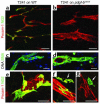
Figure 1
Figure 1
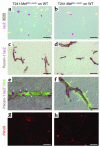
Reduced pericyte recruitment and dilated vessels in tumors transplanted into pdgf-bret/ret mice. Double staining of endothelium (Pecam-1, red) and pericytes/VSMCs (SMA or NG2, green) in the vasculature of tumors and surrounding normal tissue of WT and pdgf-bret/ret mice. Recruitment of pericytes to tumor vessels was higher in WT (a) than in pdgf-bret/ret mice (b), and vessels in tumors grown on pdgf-bret/ret mice were morphologically abnormal and significantly dilated. Vessels in the surrounding dermal tissue show continuous coverage and circular arrangement of mural cells in both WT (c) and pdgf-bret/ret mice (d). NG2 staining of the pericytes demonstrated their close association with the endothelium in tumors in WT mice (e), whereas they were partially or completely detached from the tumor endothelium in pdgf-bret/ret mice (f and g, arrows). Bars: 50 μm.
Figure 2
Figure 2
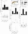
Quantitative analyses of tumor vessels in T241 tumors grown on WT C57BL6 mice (T241/WT), T241 tumors grown on pdgf-bret/ret mice (T241/ret), T241-B tumors grown on WT C57BL6 mice (T241-B/WT), and T241-B tumors grown on pdgf-bret/ret mice (T241-B/ret). (a) Mean vessel diameter. T241/WT, 11.7 ± 9; T241/ret, 26.8 ± 16; T241-B/WT, 14.5 ± 5.5; T241-B/ret, 14.9 ± 6.5 μm. (b) Scatter plot of data in a. (cf) Analysis of pericyte density and coverage. (c) Example of collected data. (d) Pericyte coverage illustrated as NG2–Pecam-1 overlapping area, expressed as a percentage of total Pecam-1–stained area. (e) Scatter plot of the data in d. (f) Pericyte density illustrated as NG2 area as a percentage of total Pecam-1 area. (gi) Pericyte “detachment” from the endothelial cells expressed as the distance (in μm) from the endothelium to the most distal part of a pericyte. (g) Example of collected data. (h) Mean distance (μm) of mural cell association with tumor endothelium. (i) Scatter plot of individual NG2-positive pericytes. *P < 0.05.
Figure 3
Figure 3
PDGF-B expression analysis. In situ hybridization revealed a similar pattern of PDGF-B expression (dark blue, arrows) by the endothelium (Pecam-1, red) of T241 tumors grown on (a) WT C57BL6 mice and (b) pdgf-bret/ret mice. PDGF-B–transfected T241 cells (T241-B cells) expressed PDGF-B both in vitro, as shown by Northern blot analysis (c), and in vivo, as shown by in situ hybridization of sections of T241-B tumors double stained for vessels (Pecam-1, red) (d). GAPDH hybridization was used to control for RNA loading. hPDGF-B, human PDGF-B. Bars: 50 μm.
Figure 4
Figure 4
Tumor cell–derived PDGF-B increases pericyte recruitment to tumor vessels. Tumor vessel endothelium is visualized by staining for endomucin (red) and pericytes by staining for NG2 (green). Low magnification shows the increased density of mural cells in central (a and b, arrows) and peripheral areas (c and d) of T241 and T241-B tumors, respectively, transplanted into WT C57BL6 mice. In parental T241 tumors, a significant proportion of the abluminal endothelial surface lacks associated pericytes (e, g, and i, arrowheads). In contrast, an almost continuous layer of pericytes is seen in T241-B tumors (f, arrows). Analysis at higher magnification reveals that the pericytes in T241-B tumors tend to circular arrangements (h), whereas the pericytes in parental T241 tumors show a more longitudinal arrangement (g). i and j show closeups of the insets in g and h. Compare also mural cell encircling of cross-sectioned vessels (insets in i and j). Bars: 50 μm (af) and 20 μm (gj).
Figure 5
Figure 5
Increased pericyte number but failure of proper pericyte investment in T241-B tumors transplanted into pdgf-bret/ret mice. Pecam-1 (red) and NG2 (green) double staining of endothelium and pericytes, respectively, reveals that PDGF-B produced by the T241-B tumors can partially rescue the defective pericyte number in pdgf-bret/ret mice (b and c, arrowheads). The pericyte density was, however, lower than that obtained after transplantation of T241-B tumors into WT mice (a). In spite of the partial rescue of pericyte number by T241-derived PDGF-B, the proper arrangement of pericytes in the vessel wall was dependent on expression of WT PDGF-B protein by the host endothelium. Note the partial or complete detachment of a majority of the pericytes in the pdgf-bret/ret mice, irrespective of whether the T241 cells express PDGF-B or not (b, and e and f, arrows), compared with T241-B tumors in WT mice (d, arrowheads). Bars: 50 μm (ac) and 20 μm (df).
Figure 6
Figure 6
Exogenous PDGF-Rβ–deficient pericytes are not recruited by tumor vessels. MEFs were isolated from XlacZ4-positive WT or PDGF-Rβ–negative embryonic day 12.5 embryos and cultured in vitro (a and b). These cultures contained similar proportions of XlacZ4-positive cells (∼10%). T241 cells mixed with MEFs at a 1:9 ratio were injected subcutaneously onto the backs of WT mice. Cells expressing lacZ (pink) were found closely associated with endothelial cells (Pecam-1, brown) in tumors mixed with PDGF-Rβ–positive MEF cells (c, arrows) but not in tumors mixed with PDGF-Rβ–negative MEF cells (d). Triple staining shows that the recruited lacZ-positive MEF cells (e, arrowheads) also express SMA (green) and that the lacZ/SMA double-positive cells are tightly associated with the endothelium (brown) (e). Vessels in tumors mixed with PDGF-Rβ–negative MEF cells lack exogenous (lacZ-positive) pericytes but recruit endogenous (lacZ-negative, SMA-positive) pericytes, as expected (f, arrows). Prelabeling of MEF cultures with Pkh26 dye indicates the presence and similar distribution of PDGF-Rβ–positive and –negative MEF cells within the tumors (g and h). Bars: 100 μm (a and b), 50 μm (c, d, g, and h), and 20 μm (e and f).
Figure 7
Figure 7
Exogenous pericyte recruitment in pdgf-bret/ret mice. Exogenous XlacZ4-positive MEF cells became associated with tumor vessels in both WT (a, arrowheads) and pdgf-bret/ret mice (b, arrowheads). Double staining for lacZ (pink) and SMA (green) revealed defective association between the exogenously recruited pericytes and the endothelium in pdgf-bret/ret tumors (d, arrow) but not in WT tumors (c, arrows). Bars: 100 μm (a and b) and 20 μm (c and d).
Figure 8
Figure 8
Model for perivascular PDGF-B protein distribution in T241 tumors and its effect on tumor vessel pericytes. WT PDGF-B protein has affinity for heparan-sulfate proteoglycans and other ECM molecules. Its expression by the endothelium is therefore expected to give rise to a depot or steep gradient of PDGF-B in the periendothelial compartment (upper left). This promotes a certain amount of pericyte recruitment, and moreover, the recruited pericytes become intimately associated with the abluminal surface of the endothelium. Additional PDGF-B protein secreted by the tumor cells (upper right) leads to additional pericyte recruitment, with retained association to the endothelium. In pdgf-bret/ret mice, the PDGF-B protein lacks the retention motif, and is therefore more freely diffusible following its release from the endothelial cells (lower left). The lower concentration or shallower gradient in the periendothelial compartment leads to reduced pericyte recruitment and defective investment of the pericytes in the microvessel wall. Additional PDGF-B protein secreted by the tumor cells (lower right) promotes the recruitment of higher numbers of pericytes, which remain abnormally associated with the endothelium.

Comment in

References

    1. Folkman J. Tumor angiogenesis: therapeutic implications. N. Engl. J. Med. 1971;285:1182–1186. - PubMed
    1. Yuan F, et al. Vascular permeability and microcirculation of gliomas and mammary carcinomas transplanted in rat and mouse cranial windows. Cancer Res. 1994;54:4564–4568. - PubMed
    1. Yuan F, et al. Vascular permeability in a human tumor xenograft: molecular size dependence and cutoff size. Cancer Res. 1995;55:3752–3756. - PubMed
    1. Hashizume H, et al. Openings between defective endothelial cells explain tumor vessel leakiness. Am. J. Pathol. 2000;156:1363–1380. - PMC - PubMed
    1. Hobbs SK, et al. Regulation of transport pathways in tumor vessels: role of tumor type and microenvironment. Proc. Natl. Acad. Sci. U. S. A. 1998;95:4607–4612. - PMC - PubMed

Publication types

MeSH terms