Case Reports
. 2003 Oct;112(8):1192-201.
doi: 10.1172/JCI18589.

A homozygous mutation in HESX1 is associated with evolving hypopituitarism due to impaired repressor-corepressor interaction

Affiliations
Case Reports

A homozygous mutation in HESX1 is associated with evolving hypopituitarism due to impaired repressor-corepressor interaction

Luciani R Carvalho et al. J Clin Invest. 2003 Oct.

Abstract

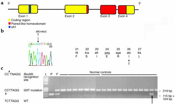
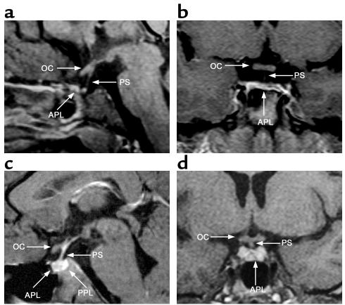
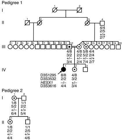
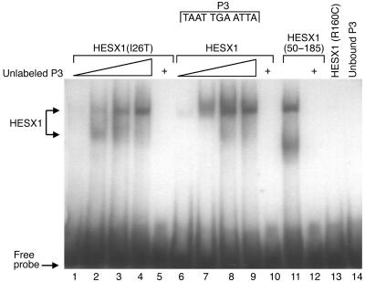
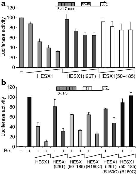
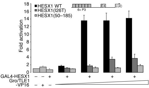
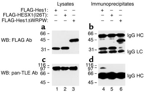
The paired-like homeobox gene expressed in embryonic stem cells Hesx1/HESX1 encodes a developmental repressor and is expressed in early development in a region fated to form the forebrain, with subsequent localization to Rathke's pouch, the primordium of the anterior pituitary gland. Mutations within the gene have been associated with septo-optic dysplasia, a constellation of phenotypes including eye, forebrain, and pituitary abnormalities, or milder degrees of hypopituitarism. We identified a novel homozygous nonconservative missense mutation (I26T) in the critical Engrailed homology repressor domain (eh1) of HESX1, the first, to our knowledge, to be described in humans, in a girl with evolving combined pituitary hormone deficiency born to consanguineous parents. Neuroimaging revealed a thin pituitary stalk with anterior pituitary hypoplasia and an ectopic posterior pituitary, but no midline or optic nerve abnormalities. This I26T mutation did not affect the DNA-binding ability of HESX1 but led to an impaired ability to recruit the mammalian Groucho homolog/Transducin-like enhancer of split-1 (Gro/TLE1), a crucial corepressor for HESX1, thereby leading to partial loss of repression. Thus, the novel pituitary phenotype highlighted here appears to be a specific consequence of the inability of HESX1 to recruit Groucho-related corepressors, suggesting that other molecular mechanisms govern HESX1 function in the forebrain.


Figures

Figure 1
Figure 1
Schematic of the HESX1 gene, HESX1(I26T) mutation, and Bsu36I restriction digest assay within a normal Brazilian population. (a) Genomic and protein structure of HESX1. HESX1 consists of four exons encoding a 185-AA protein. The engrailed homology domain (AAs 21–27) is encoded by exon 1 and the prd-like homeodomain by exons 2–4. (b) Homozygous missense mutation within exon 1 leads to the substitution of an isoleucine residue (codon 26) by threonine in the engrailed homology domain eh1. (c) Bsu36I restriction digests in pedigree 1 (F, father; P, patient) and 18 control samples. Digestion of a homozygous sample results in one band (104 + 115 bp fragment size), digestion of a heterozygous sample results in two bands (a 219-bp band from the normal allele and a band with the 104-bp and 115-bp fragments from the mutant allele), and digestion of a homozygous wild-type sample results in a single 219-bp band. The arrow indicates a normal control who is heterozygous for HESX1(I26T).
Figure 2
Figure 2
Neuroimaging studies of the patient with the HESX1(I26T) mutation (a and b) and a normal subject (c and d). (a) Sagittal MRI scan of the patient homozygous for HESX1(I26T) at 21 years, showing a thin, continuous pituitary stalk (PS), a normal optic chiasm (OC), and a severely hypoplastic anterior pituitary lobe (APL). (b) Coronal MRI scan of the patient at 11 years, showing a hypoplastic anterior pituitary lobe and a normal optic chiasm, with a thin pituitary stalk that is not clearly visualized. (c) Normal sagittal MRI scan showing a normal anterior pituitary lobe, a normal pituitary stalk, a normally sited PPL, and a normal optic chiasm. (d) Normal coronal MRI scan showing a normal anterior pituitary lobe and a normal optic chiasm with a normal pituitary stalk.
Figure 3
Figure 3
Segregation analysis of the I26T mutation within pedigree 1 (the family of the index patient with CPHD) and pedigree 2 (the normal control family) using the highly polymorphic HESX1-flanking markers D3S1295, D3S3532, and D3S3616. The figure shows the presence of haplotype 8, 2, 4 linked to the HESX1(I26T) mutation in individuals from pedigree 1 (III-12, III-13, III-14, IV-1, and IV-2) and pedigree 2 (I-1 and II-1).
Figure 4
Figure 4
Electrophoretic mobility shift assay. Increasing amounts of in vitro–translated HESX1(I26T) (1 μl, 5 μl, 10 μl, and 20 μl) were added to the consensus palindromic DNA-binding site P3 (lanes 1–4). Identical amounts of wild-type HESX1 were added to P3 (lanes 6–9). Lane 11 shows addition of the highest amount of HESX1(50–185) (20 μl) added to P3. Lanes 5, 10, and 12 show the highest amounts (20 μl) of the respective protein construct [lane 5, HESX1(I26T); lane 10, wild-type HESX1; lane 12, HESX1(50–185)] added to radiolabeled P3 and excess (15 pM) unlabeled P3. Lane 13 represents the addition of 20 μl HESX1(R160C) to P3, while lane 14 shows the effect of free probe in the absence of protein. Plus signs at the top of lanes 5, 10, and 12 indicate the addition of unlabeled P3.
Figure 5
Figure 5
(a) HESX1(I26T) leads to impaired repression at the SV40 promoter. Increasing concentrations of GAL4-HESX1, GAL4-HESX1(I26T), and GAL4-HESX1(50–185) (10 ng, 100 ng, 250 ng, and 500 ng) were cotransfected with the SV40 promoter reporter construct. GAL4-HESX1 led to a dose-dependent repression of the SV40 promoter (67.5% at the highest concentration tested), whereas GAL4-HESX1(I26T) was associated with impaired repression (35.2% at the highest concentration tested) (P = 0.02). GAL4-HESX1(50–185) was associated with 24.4% repression, which did not achieve statistical significance (P = 0.19). The results represent the means of three independent experiments, each performed in triplicate. (b) HESX1(I26T) leads to impaired repression of Bix activation. Two different concentrations (10 ng and 250 ng) of GAL4-HESX1, GAL4-HESX1(I26T), GAL4-HESX1(50–185), GAL4-HESX1(R160C), GAL4-HESX1(I26T)(R160C), and GAL4-HESX1(50–185)(R160C) were cotransfected with a reporter containing six paired-class binding sites upstream of the E4 promoter that drives luciferase expression [(P3)6E4], and with the paired-class activator Bix. GAL4-HESX1 was associated with a tenfold repression of Bix activation at the highest concentration tested, whereas both GAL4-HESX1(I26T) and GAL4-HESX1(50–185) led to a threefold repression of Bix activation at the (P3)6E4 promoter. GAL4-HESX1(R160C) was associated with a fourfold repression of Bix activation, whereas the introduction of the I26T mutation [GAL4-HESX1(I26T)(R160C)] or the removal of the N-terminal AAs [GAL4-HESX1(50–185)(R160C)] led to a twofold repression and no repression of Bix activation, respectively. A constant concentration of Bix was used in all experiments (250 ng). The results represent the means of three independent experiments, each performed in triplicate.
Figure 6
Figure 6
HESX1(I26T) shows impaired interaction with Gro/TLE-VP16 in transient transfection assays. A constant concentration of GAL4-HESX1, GAL4-HESX1(I26T), or GAL4-HESX1(50–185) (250 ng) was cotransfected with increasing concentrations of Gro/TLE-VP16 (10 ng, 100 ng, 400 ng, and 800 ng) together with the (P3)E4 reporter construct. GAL4-HESX1 led to a 14-fold activation of (P3)E4 reporter activity when cotransfected with the highest concentration of VP16-Gro/TLE, whereas GAL4-HESX1(I26T) was associated with a threefold activation of the reporter. GAL4-HESX1(50–185) did not lead to any activation of the reporter above base line. Control transfections with each of the expression constructs transfected individually show that, separately, neither GAL4-HESX1 nor Gro/TLE-VP16 can bind and activate the (P3)E4 reporter. The results represent the means of three independent experiments, each performed in triplicate.
Figure 7
Figure 7
Interaction of HESX1 with Gro/TLE. HEK 293 cells were transfected with plasmids encoding FLAG epitope–tagged HESX1 (lanes 1 and 4), HESX1(I26T) (lanes 2 and 5), or a truncated form of the bHLH protein Hes1, lacking the C-terminal WRPW motif (lanes 3 and 6). (b and d) Cell lysates were prepared and subjected to immunoprecipitation with anti–FLAG epitope antibodies, followed by SDS-PAGE. (a and c) One-eighth of each input lysate, collected prior to incubation with antibodies, was also subjected to gel electrophoresis. After transfer to nitrocellulose, Western blotting (WB) was performed with either anti-FLAG (a and b) or anti–Gro/TLE (pan-TLE; c and d) antibodies. Both HESX1 and HESX1(I26T) migrated as roughly 32-kDa proteins, but only HESX1 coimmunoprecipitated with endogenous Gro/TLE proteins of roughly 95 kDa. The specificity of this interaction was demonstrated further by the finding that Hes1ΔWRPW also failed to interact with Gro/TLE, as previously shown (22). Positions of migration of the IgG heavy chain (HC) and light chain (LC) are indicated. Positions of size standards are indicated in kilodaltons.

References

    1. Schwind JL. The development of the hypophysis cerebri of the albino rat. Am. J. Anat. 1928;41:295–315.
    1. Kaufman, M.H. 1992. An atlas of mouse development. Academic Press, Elsevier Science. Oxford, United Kingdom. 39–210.
    1. Dattani MT, et al. Mutations in the homeobox gene HESX1/Hesx1 associated with septo-optic dysplasia in human and mouse. Nat. Genet. 1998;19:125–133. - PubMed
    1. Martinez-Barbera JP, Rodriguez TA, Beddington RS. The homeobox gene Hesx1 is required in the anterior neural ectoderm for normal forebrain formation. Dev. Biol. 2000;223:422–430. - PubMed
    1. Thomas PQ, et al. Heterozygous HESX1 mutations associated with isolated congenital pituitary hypoplasia and septo-optic dysplasia. Hum. Mol. Genet. 2001;10:39–45. - PubMed

Publication types