. 2003 Oct 15;552(Pt 2):525-33.
doi: 10.1111/j.1469-7793.2003.00525.x.

Membrane properties of type II spiral ganglion neurones identified in a neonatal rat cochlear slice

Affiliations

Membrane properties of type II spiral ganglion neurones identified in a neonatal rat cochlear slice

Daniel J Jagger et al. J Physiol. .

Abstract

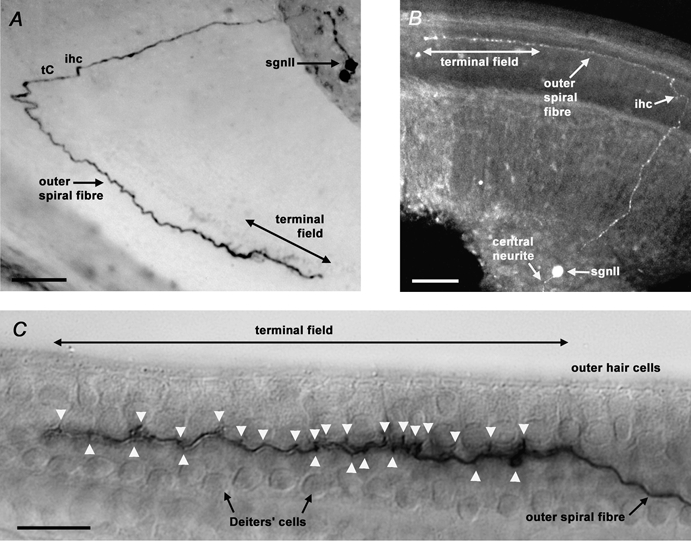
Neuro-anatomical studies in the mammalian cochlea have previously identified a subpopulation of approximately 5 % of primary auditory neurones, designated type II spiral ganglion neurones (sgnII). These neurones project to outer hair cells and their supporting cells, within the 'cochlear amplifier' region. Physiological characterization of sgnII has proven elusive. Whole-cell patch clamp of spiral ganglion neurones in P7-P10 rat cochlear slices provided functional characterization of sgnII, identified by biocytin or Lucifer yellow labelling of their peripheral neurite projections (outer spiral fibres) subsequent to electrophysiological characterisation. SgnII terminal fields comprised multiple outer hair cells and supporting cells, located up to 370 mum basal to their soma. SgnII firing properties were defined by rapidly inactivating A-type-like potassium currents that suppress burst firing of action potentials. Type I spiral ganglion neurones (sgnI), had shorter radial projections to single inner hair cells and exhibited larger potassium currents with faster activation and slower inactivation kinetics, compatible with the high temporal firing fidelity seen in auditory nerve coding. Based on these findings, sgnII may be identified in future by the A-type current. Glutamate-gated somatic currents in sgnII were more potentiated by cyclothiazide than those in sgnI, suggesting differential AMPA receptor expression. ATP-activated desensitising inward currents were comparable in sgn II and sgnI. These data support a role for sgnII in providing integrated afferent feedback from the cochlear amplifier.


Figures

Figure 1
Figure 1. Biocytin and Lucifer yellow labelling confirmed the identity of type II spiral ganglion neurones by resolving their outer spiral fibres
A, extended-focus view of a biocytin-filled type II spiral ganglion neurone (sgnII) in the mid-apical region of a P8 slice, revealing an outer spiral fibre travelling 340 μm in the basal direction from the inner hair cell (ihc) region and the tunnel of Corti (tC). Ten outer hair cells (ohc) in rows 1 and 2 were innervated within the terminal field. The other cell labelled in the ganglion had type I neurone-like (sgnI) properties, but had no neurites evident. Scale bar = 50 μm. B, confocal optical section reconstruction of a Lucifer yellow-filled sgnII in the apical region of a P7 slice. Outer spiral fibre length was 310 μm, with 13 ohc contacted in row 1. Scale bar = 50 μm. C, lateral view of the neurite terminal field of a biocytin-filled P9 apical sgnII. Terminals (arrowheads) appear to be onto ohc and Deiters’ cells. 16 ohc in row 1 were innervated by this neurone. Scale bar = 20 μm
Figure 2
Figure 2. Biocytin labelling confirmed the identity of type I spiral ganglion neurones by resolving their radial fibres
Extended-focus view of a biocytin-filled type I spiral ganglion neurone (sgnI) in the basal region of a P8 slice, revealing a radial fibre innervating a single inner hair cell (ihc). Scale bar = 50 μm. Inset, detail of sgnI dendrite, showing terminal contacts on the modiolar face and the basal pole of the ihc. Scale bar = 20 μm.
Figure 3
Figure 3. Type II spiral ganglion neurones displayed dominant inactivating outward currents
A, rapidly inactivating outward currents in type II spiral ganglion neurone (sgnII; left panel, marked *), and outward currents dominated by a slower inactivation time constant in type I spiral ganglion neurone (sgnI; right panel). Transient inward sodium currents are evident at the start of the depolarizing voltage steps, and slowly activating inward currents were evident during hyperpolarizing voltage steps in both cells. B, voltage-dependent activation kinetics of the outward currents in identified sgnII (▪, n = 8) and identified sgnI (○, n = 17). In both cell types the time to peak current decreased with increasing depolarisation. The curves are single exponential fits to the mean data, with 32 mV per efold increase in activation time for sgnII and 80 mV for sgnI.C, relative amplitudes of peak and steady-state (s-s) currents in identified sgnII and sgnI. Currents measured at beginning (peak) and end (s-s) of 200 ms test potentials to +40 mV (holding potential -100 mV). D, outward currents of identified sgnII had shorter primary inactivation time constants (▪) than those of identified sgnI (•). Non-traced neurones with sgnII-like electrical phenotype (□), and sgnI-like electrical phenotype (○) are included for comparison. E, primary inactivation time constants of sgnII and sgnI plotted as a percentage of total inactivation. SgnII currents were dominated by a short primary time constant ( < 55 ms, 64-80 % contribution), whereas sgnI current inactivation showed longer and less dominant primary time constants (>60 ms, 22-69 % contribution). The data in D and E were determined during 2000 ms depolarising steps to +40 mV, following -100 mV conditioning (1000 ms). F, voltage dependence of activation (▪) and steady-state inactivation (□) for A-type currents in the same sgnII shown in A. A-type currents derived by subtraction (see Methods). Activation threshold was close to -50 mV. Activation and inactivation voltage dependence were described by single modified Boltzmann relationships (continuous lines), with half-maximal activation (V0.5) estimated at -12.2 mV (slope factor, k = 12.0 mV per efold change in conductance), and half-maximal inactivation (V0.5) estimated at -76.4 mV (k = 20.0). G, bath-applied 5 mM 4-aminopyridine (4AP) selectively blocked the A-type potassium current in sgnII (2000 ms voltage step to +40 mV, from 1000 ms conditioning potential at -100 mV). In the presence of 4AP, only a ‘residual’ fast-activating sustained outward current remained.
Figure 4
Figure 4. Outward current inactivation shaped the type II spiral ganglion neurone voltage response
A, under current clamp conditions, comparison of voltage responses of the type II spiral ganglion neurone (sgnII; left panel) and type I spiral ganglion neurone (sgnI; right panel) demonstrated that sgnII more rapidly recruit input resistance during square wave current injections (100-700 pA) at the resting potential. This was consistent with the inactivation of the dominant outward currents in sgnII. Resting potentials of -64 mV (sgnII) and -77 mV (sgnI) are offset for clarity. B, steady-state current-voltage relationships for groups of sgnII (▪; n = 4 cells) and sgnI (•; n = 8 cells). SgnII showed a steeper slope around the resting potential reflecting their higher input resistance despite lower resting potentials. SgnI showed a pronounced rectification attributable to their greater steady-state voltage-dependent conductance.

References

    1. Adamson CL, Reid MA, Mo ZL, Bowne-English, Davis RL. Firing features and potassium channel content of murine spiral ganglion neurons vary with cochlear location. J Comp Neurol. 2002;447:331–350. - PubMed
    1. Ashmore JF, Kolston PJ. Hair cell based amplification in the cochlea. Curr Op Neurobiol. 1994;4:503–508. - PubMed
    1. Berglund AM, Brown MC. Central trajectories of type II spiral ganglion cells from various cochlear regions in mice. Hear Res. 1994;75:121–130. - PubMed
    1. Berglund AM, Ryugo DK. Hair cell innervation by spiral ganglion neurons in the mouse. J Comp Neurol. 1987;255:560–570. - PubMed
    1. Brown MC. Morphology of labeled afferent fibers in the guinea pig cochlea. J Comp Neurol. 1987;260:591–604. - PubMed

Publication types

LinkOut - more resources