. 2003 Oct 15;23(28):9418-27.
doi: 10.1523/JNEUROSCI.23-28-09418.2003.

Histone deacetylase inhibition by sodium butyrate chemotherapy ameliorates the neurodegenerative phenotype in Huntington's disease mice

Affiliations

Histone deacetylase inhibition by sodium butyrate chemotherapy ameliorates the neurodegenerative phenotype in Huntington's disease mice

Robert J Ferrante et al. J Neurosci. .

Abstract

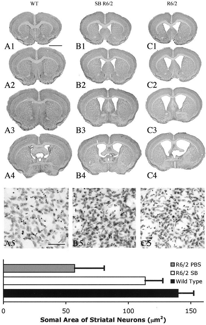
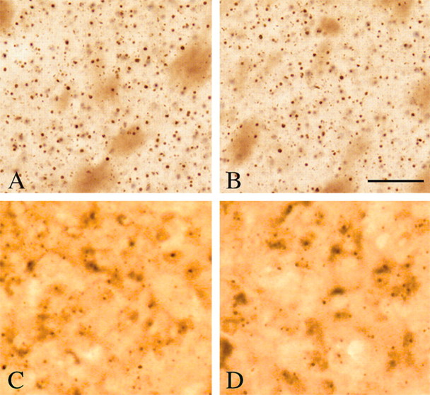
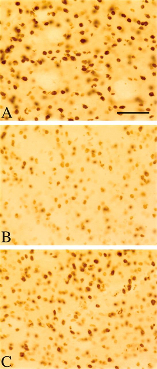
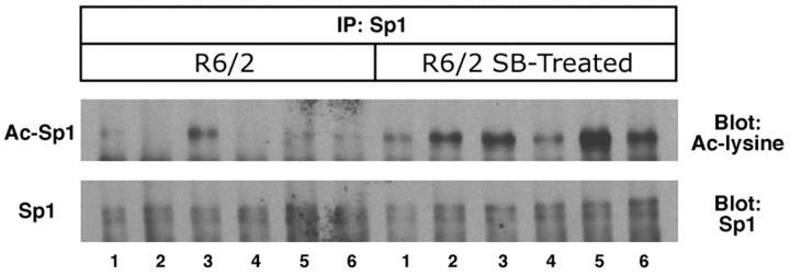
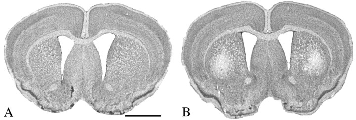
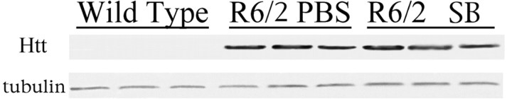
The precise cause of neuronal death in Huntington's disease (HD) is unknown. Although no single specific protein-protein interaction of mutant huntingtin has emerged as the pathologic trigger, transcriptional dysfunction may contribute to the neurodegeneration observed in HD. Pharmacological treatment using the histone deacetylase inhibitor sodium butyrate to modulate transcription significantly extended survival in a dose-dependent manner, improved body weight and motor performance, and delayed the neuropathological sequelae in the R6/2 transgenic mouse model of HD. Sodium butyrate also increased histone and Specificity protein-1 acetylation and protected against 3-nitropropionic acid neurotoxicity. Microarray analysis showed increased expression of alpha- and beta-globins and MAP kinase phosphatase-1 in sodium butyrate-treated R6/2 mice, indicative of improved oxidative phosphorylation and transcriptional regulation. These findings strengthen the hypothesis that transcriptional dysfunction plays a role in the pathogenesis of HD and suggest that therapies aimed at modulating transcription may target early pathological events and provide clinical benefits to HD patients.


Figures

Figure 1.
Figure 1.
Survival, body weight, and motor performance analysis in sodium butyrate (SB)-treated R6/2 mice. Cohorts of R6/2 mice (n = 20) were treated from day 21 with an intraperitoneal injection of 100, 200, 400, 600, 1200, 5000, and 10,000 mg · kg-1 · d-1 sodium butyrate. Kaplan-Meier probability of survival analyses of sodium butyrate treatments in R6/2 mice and PBS-treated R6/2 mice are presented in A. Survival was significantly extended in all sodium butyrate dosing paradigms except the 100 mg · kg-1 · d-1 dose, with the greatest increase at the 1200 mg · kg-1 · d-1 dose. The data for the 5000 and 10,000 mg · kg-1 · d-1 doses resulted in marked rapidly coursing morbidity and mortality at on set of treatment and are not included. Effects of intraperitoneal sodium butyrate treatment on body weight in R6/2 HD transgenic mice are shown in B. A significant reduction in body weight loss was observed only after 12 weeks of age. Effects of intraperitoneal sodium butyrate treatment on rotarod performance (C) significantly improved motor performance in R6/2 HD transgenic mice throughout the temporal sequence of the experiment at each of the doses tested. *p < 0.01; **p < 0.001.
Figure 2.
Figure 2.
Gross brain and histopathological neuroprotection with sodium butyrate (SB) treatment. Photomicrographs of coronal serial step sections from the rostral neostriatum through the level of the anterior commissure in a wild-type littermate mouse (A1-A4), a sodium butyrate-treated (1.2 gm · kg-1 · d-1) R6/2 HD transgenic mouse (B1-B4), and a PBS-treated (C1-C4) R6/2 HD transgenic mouse at 90 d are shown. There was gross atrophy of the brain in the PBS-treated R6/2 mouse along with ventricular hypertrophy (C1-C4) compared with the wild-type littermate control mouse (A1-A4). In contrast, the sodium butyrate-treated R6/2 mouse brain (B1-B4) showed reduced gross brain atrophy and ventricular enlargement compared with the PBS-treated R6/2 mouse (C1-C4). Corresponding Nissl-stained tissue sections from the dorsomedial aspect of the neostriatum in a wild-type littermate control (A5), sodium butyrate-treated R6/2 mouse (B5), and PBS-treated R6/2 mouse (C5) are also shown. There was marked neuronal atrophy in the PBS-treated R6/2 mouse, with significantly less neuronal atrophy (p < 0.01) in the sodium butyrate-treated R6/2 mouse compared with this PBS-treated R6/2 mouse. The histogram shows means and SDs of somal areas of striatal neuronsquantitated in each group of mice (n = 10) (see Materials and Methods). Scale bars: A1-A4, B1-B4, C1-C4, 2 mm; A5, B5, C5, 100 μm.
Figure 3.
Figure 3.
Huntingtin and ubiquitin immunoreactivity in sodium butyrate-treated R6/2 mice. Huntingtin immunostained tissue sections from the neostriatum of a PBS-treated R6/2 transgenic mouse (A) and sodium butyrate-treated R6/2 HD transgenic mouse (B) euthanized at 90 d of age are shown. Sodium butyrate (1.2 gm · kg-1 · d-1) and PBS treatments were started at 21 d. There were no significant differences (p < 0.27) in the number and size of huntingtin aggregates and inclusions between treated and untreated mice. Similarly, no differences were observed in ubiquitin-positive inclusions between PBS-treated and sodium butyrate-treated R6/2 mice (C and D, respectively). Scale bar: (in B) A-D, 100 μm.
Figure 4.
Figure 4.
Western blot of acetylated H3 and H4 in sodium butyrate (sb)-treated R6/2 mice. R6/2 mice were treated with 1.2 gm · kg-1 · d-1 sodium butyrate for 2 weeks starting at 42 d and euthanized at 56 d. Hypoacetylation of H3 and H4 immunoreactivities were present in R6/2 mice compared with wild-type littermate control mice (WT). Sodium butyrate-treated R6/2 mice showed a marked increase in acetylated H3 and H4 activity. Protein levels were determined using Coomassie protein assay.
Figure 5.
Figure 5.
Striatal tissue immunohistochemistry of acetylated histone 4 in sodium butyrate-treated R6/2 mice. At 90 d of age, robust acetylated histone 4 immunohistochemistry was present in wild-type littermate control striatal tissue specimens (A), with hypoacetylation in the R6/2 mice (B). C, Sodium butyrate treatment (1.2 gm · kg-1 · d-1) increased acetylation of histone 4 in R6/2 mice. Scale bar: (in A) A-C, 100 μm.
Figure 6.
Figure 6.
The histone deacetylase inhibitor sodium butyrate (SB) enhances Sp1 acetylation in vivo. Cohorts of R6/2 mice (n = 6) were treated daily with sodium butyrate (1.2 gm/kg) or PBS intraperitoneal injections for 2 weeks. Sp1 acetylation levels from homogenized brains of PBS- and sodium butyrate-treated R6/2 mice were determined by immunoprecipitation (IP) using an Sp1 antibody followed by immunoblotting using acetyl lysine (Ac-lysine) antibody (Ac-Sp1) or Sp1 antibody alone (Sp1). Sp1 acetylation was increased in sodium butyrate-treated R6/2 mice. Note that levels of Sp1 did not change with sodium butyrate treatment.
Figure 7.
Figure 7.
Sodium butyrate neuroprotection from 3-NP acid toxicity in R6/2 mice. Groups of R6/2 mice (n = 10) were treated for 2 weeks with 1.2 gm · kg-1 · d-1 sodium butyrate or PBS starting at 42 d. 3-NP was administered at the beginning of the second week for 4.5 d along with PBS and sodium butyrate treatments. Sodium butyrate treatment prevented 3-NP striatal-induced damage in R6/2 mice (A) compared with PBS-treated R6/2 mice (B). Histopathological evaluation of 3-NP-induced striatal lesions showed bilateral striatal lesions (areas of pallor) in PBS-treated R6/2 mice (B). Scale bar: (in A) A, B, 2 mm.
Figure 8.
Figure 8.
Huntingtin (Htt) expression in sodium butyrate (SB)-treated R6/2 mice. Transgene expression was determined in whole-brain samples by Western blot analysis of sodium butyrate-treated (1.2 gm · kg-1 · d-1) R6/2 mice, PBS-treated R6/2 mice, and wild-type mice. No differences in huntingtin expression levels in sodium butyrate-treated R6/2 mice were observed compared with PBS-treated mice. Parallel blots probed against α-tubulin were run to normalize for gel loading.
Figure 9.
Figure 9.
MKP-1 is increased by sodium butyrate treatment. Northern blot analysis of 2 μg of total RNA from brain samples of sodium butyrate-treated (1.2 gm · kg-1 · d-1) and untreated R6/2 mice (n = 4 in each group) confirmed that sodium butyrate treatment increases the expression of MKP-1 mRNA compared with both untreated R6/2 mice and wild-type (WT) littermate control mice. A β-actin hybridization signal on the same blot (shown) was used as a loading control. Quantitative and statistical data are presented in Table 2.

References

    1. Alston TA, Mela L, Bright HJ ( 1977) 3-Nitropropionate, the toxic substance of Indigofera, is a suicide inactivator of succinate dehydrogenase. Proc Natl Acad Sci USA 74: 3767-3771. - PMC - PubMed
    1. Andreassen OA, Ferrante RJ, Huang HM, Dedeoglu A, Park L, Ferrante KL, Kwon J, Borchelt DR, Ross CA, Gibson GE, Beal MF ( 2001a) Dichloroacetate exerts therapeutic effects in transgenic mouse models of Huntington's disease. Ann Neurol 50: 112-117. - PubMed
    1. Andreassen OA, Ferrante RJ, Dedeoglu A, Beal MF ( 2001b) Lipoic acid improves survival in transgenic mouse models of Huntington's disease. NeuroReport 12: 3371-3374. - PubMed
    1. Beal MF ( 2000) Energetics in the pathogenesis of neurodegenerative diseases. Trends Neurosci 7: 298-304. - PubMed
    1. Beal MF, Brouillet E, Jenkins BG, Ferrante RJ, Kowall NW, Miller JM, Storey E, Srivastava R, Rosen BR, Hyman BT ( 1993) Neurochemical and histo-logic characterization of striatal excitotoxic lesions produced by the mitochondrial toxin 3-nitropropionic acid. J Neurosci 13: 4181-4192. - PMC - PubMed

Publication types

MeSH terms