. 2003 Oct 28;100(22):12889-94.
doi: 10.1073/pnas.1735556100. Epub 2003 Oct 15.

Early phagosomes in dendritic cells form a cellular compartment sufficient for cross presentation of exogenous antigens

Affiliations

Early phagosomes in dendritic cells form a cellular compartment sufficient for cross presentation of exogenous antigens

Anne L Ackerman et al. Proc Natl Acad Sci U S A. .

Abstract

Conventionally, MHC class I-restricted antigen (Ag) processing requires the action of the multimolecular peptide-loading complex within the endoplasmic reticulum (ER). Here we show that early phagosomes from human dendritic cells (DCs) contain the peptide-loading complex, incorporating MHC class I, beta2 microglobulin, transporter associated with Ag processing (TAP), calreticulin, tapasin, and ERp57. Antigenic peptides could be translocated into purified phagosomes by TAP and loaded onto cognate class I molecules, inducing their specific dissociation from the loading complex. Endoglycosidase H-sensitive class I molecules were detected at the DC cell surface, suggesting that these molecules traffic there directly from phagosomes. Macropinocytosis also allowed internalized soluble Ags access to an ER-like compartment containing the class I loading complex. Blockade of TAP by endocytosis of a soluble derivative of human cytomegalovirus protein US6 confirmed that, although retrotranslocation into the cytosol is critical for processing, efficient association of class I molecules with peptides derived from exogenous Ags occurs within a compartment directly accessible to internalized proteins. Together, this evidence suggests that early phagosomes and pinosomes facilitate cross presentation of exogenous Ags by DCs.


Figures

Fig. 1.
Fig. 1.
DC phagosomes contain a properly assembled MHC class I loading complex. Digitonin lysates of whole cells (TCL), total phagosomes (TP), TAP immunoprecipitates from phagosome lysates (TAP), or supernatants from the TAP immunoprecipitates (NTA), were separated by SDS/PAGE and immunoblotted for MHC class I heavy chains (A), CRT (B), TAP (C), ERAP (D), L22 ribosomal protein (E), tapasin (F), and ERp57 (G). (A) Samples were treated where indicated with endoH. (E and F) Cells were pretreated with N-ethylmaleimide before lysis or phagosome preparation, and the samples were reduced where indicated with DTT. Cont, isotype control immunoprecipitates. (C) A second TAP immunoprecipitate of the phagosomal lysate (20 TAP) confirms TAP depletion. Mr markers are indicated on the left.
Fig. 2.
Fig. 2.
TAP in phagosomes facilitates peptide transport and MHC class I peptide loading. (A) Phagosomes purified from KG-1 DLCs were incubated with a radioiodinated glycosylation acceptor peptide for the indicated times. Glycosylated peptides were recovered by using Con A-Sepharose beads and counted in a γ-counter (□). Treatment with 15 μM ICP47 (•) abrogated transport to levels seen in apyrase-treated negative controls (gray line). (B) Phagosomes purified from HLA-A3+ immature DCs were incubated with radioiodinated HLA-A3-binding peptide, [125I]nef7B, for the indicated times. HLA-A3 (□), and HLA-B,C (•) molecules were immunoprecipitated, and the quantity of bound peptide was counted in a γ-counter. Bound peptide recovered in the presence of apyrase (gray line) served as a negative control.
Fig. 3.
Fig. 3.
Peptide loading induces MHC class I dissociation from the loading complex in phagosomes. (A) Phagosomes from [35S]methionine-labeled KG-1.Kb DLCs were incubated with either SIINFEKL (S) or nef7B (N) peptide for the indicated times. TAP immunoprecipitation shows that SIINFEKL specifically induces dissociation of Kb but not HLA molecules. Y3 (Kb) and 4E (HLA-B,C) immunoprecipitates served as mobility controls. (B) [35S]Methionine-labeled immature DCs incubated for 30 min with or without latex beads were surface biotinylated. After lysis, cell surface MHC class I and tapasin molecules were isolated with streptavidin beads. After stripping with SDS, MHC class I heavy chains were reprecipitated and incubated with or without Endo H. MHC class I and tapasin served as mobility controls.
Fig. 4.
Fig. 4.
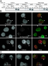
Internalized exogenous Ags can access an ER-like compartment. (A) Internalized anti-tapasin IgG can interact with tapasin–MHC class I complexes. [35S]Methionine-labeled KG-1 DLCs were allowed to internalize soluble PaSta1 or control GAP.A3 mAbs or PaSta1-coated latex beads. After lysis in digitonin, Abs were isolated with protein G-Sepharose. SDS eluates were reprecipitated with Abs against either human MHC class I (3B10.7) or a control Ab (Y3) and treated with or without endoH. An MHC class I (w6/32) immunoprecipitate served as a mobility control. (B) Internalized Ags colocalize with ER components early after endocytosis. KG-1 DLCs were allowed to internalize OVA (Top), anti-tapasin IgG (PaSta1, Middle), and US6 (Bottom) for 10 min and then fixed and stained for calnexin by using AF8 (Top and Bottom) or R.CNX (Middle). Internalized proteins colocalized with calnexin in peripheral vesicles (white arrowheads). (C) OVA was internalized by KG-1 DLCs for 10 min and then removed by extensive washing. Cells were incubated for an additional 30 min at 37°C and then fixed and stained for the lysosomal marker CD63 (H5C6). CD63 colocalized with internalized OVA in internal vesicles (white arrowheads).
Fig. 5.
Fig. 5.
Soluble US6 interacts with TAP to down-regulate surface MHC class I expression. The association of US6 with TAP in intact cells was shown by immunoprecipitation of internalized US6 (A) and TAP (B) from immature DCs and blotting for associated TAP and US6, respectively. Although both are present in both whole cells and phagosomes (lanes 1 and 2), the two molecules associate only in US6-treated cells (indicated by +, lane 5). Sample designations are as in Fig. 1. Treatment with US6 down-regulated surface levels of MHC class I in immature DCs (C) and KG-1 DLCs (D) in a dose-dependent manner (E). Cells were treated with the indicated amounts of soluble US6 for 16hat37°C and then analyzed by flow cytometry for MHC class I expression. Dark lines represent MHC class I surface expression in treated samples, whereas thin lines display untreated controls. The percentage of down-regulation is quantitated in E for DCs (•) and KG-1 (♦).
Fig. 6.
Fig. 6.
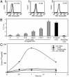
Soluble US6 inhibits the cross presentation of exogenous Ags. (A) KG1.Kb cells were incubated for 16 h in 10 mg/ml OVA (bold lines) or BSA (thin lines) with the indicated concentrations of soluble US6. SIINFEKL Kb complexes were visualized by flow cytometry by using the 25D1.16 mAb. (B) Inhibition was confirmed by coculture of the SIINFEKL-Kb-specific T hybridoma, B3Z, with KG-1.Kb cells pretreated with OVA, and soluble US6. IL-2 production was quantified by IL-2 ELISA. (C) Binding to MHC class I of a peptide precursor requiring processing is inhibited by both lactacystin and soluble US6. An N- and C-terminally extended, radioiodinated polypeptide containing the embedded nef7B (A3-binding) epitope was incubated with HLA-A3+ immature DCs for the indicated times before extraction and immunoprecipitation. Bound peptide was quantitated by counting in a γ-counter.

References

    1. Lankat-Buttgereit, B. & Tampe, R. (2002) Physiol. Rev. 82 187–204. - PubMed
    1. Radcliffe, C. M., Diedrich, G., Harvey, D. J., Dwek, R. A., Cresswell, P. & Rudd, P. M. (2002) J. Biol. Chem. 277 46415–46423. - PubMed
    1. Dick, T. P., Bangia, N., Peaper, D. R. & Cresswell, P. (2002) Immunity 16 87–98. - PubMed
    1. Serwold, T., Gonzalez, F., Kim, J., Jacob, R. & Shastri, N. (2002) Nature 419 480–483. - PubMed
    1. Saric, T., Chang, S. C., Hattori, A., York, I. A., Markant, S., Rock, K. L., Tsujimoto, M. & Goldberg, A. L. (2002) Nat. Immunol. 3 1169–1176. - PubMed

Publication types

MeSH terms