. 2003 Oct 28;100(22):13042-7.
doi: 10.1073/pnas.2135111100. Epub 2003 Oct 15.

Interaction between the Alzheimer's survival peptide humanin and insulin-like growth factor-binding protein 3 regulates cell survival and apoptosis

Affiliations

Interaction between the Alzheimer's survival peptide humanin and insulin-like growth factor-binding protein 3 regulates cell survival and apoptosis

Maaria Ikonen et al. Proc Natl Acad Sci U S A. .

Abstract

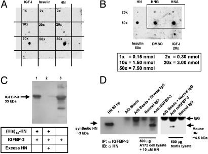
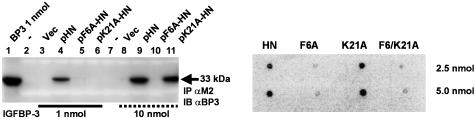
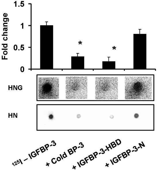
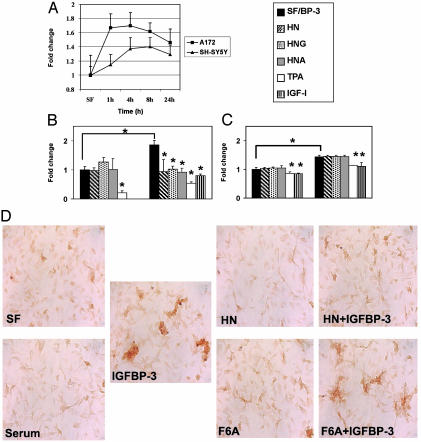
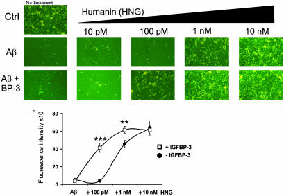
Insulin-like growth factor-binding protein-3 (IGFBP-3) regulates IGF bioactivity and also independently modulates cell growth and survival. By using a yeast two-hybrid screen to identify IGFBP-3-interacting proteins, we cloned humanin (HN) as an IGFBP-3-binding partner. HN is a 24-aa peptide that has been shown to specifically inhibit neuronal cell death induced by familial Alzheimer's disease mutant genes and amyloid-beta (Abeta). The physical interaction of HN with IGFBP-3 was determined to be of high affinity and specificity and was confirmed by yeast mating, displaceable pull-down experiments with (His)-6-tagged HN, and ligand blot experiments. Co-immunoprecipitation of IGFBP-3 and HN from mouse testes confirmed the interaction in vivo. In cross-linking experiments, HN bound IGFBP-3 but did not compete with IGF-I-IGFBP-3 binding; competitive ligand dot blot experiments revealed the 18-aa heparin-binding domain of IGFBP-3 as the binding site for HN. Alanine scanning determined that F6A-HN mutant does not bind IGFBP-3. HN but not F6A-HN inhibited IGFBP-3-induced apoptosis in human glioblastoma-A172. In contrast, HN did not suppress IGFBP-3 response in SH-SY5Y neuroblastoma and mouse cortical primary neurons. In primary neurons, IGFBP-3 markedly potentiated HN rescue ability from Abeta1-43 toxicity. In summary, we have identified an interaction between the survival peptide HN and IGFBP-3 that is pleiotrophic in nature and is capable of both synergistic and antagonistic interaction. This interaction may prove to be important in neurological disease processes and could provide important targets for drug development.


Figures

Fig. 1.
Fig. 1.
IGFBP-3 binds HN. (A) A ligand dot blot experiment showing IGFBP-3 binding to HN. HN, IGF-I, and insulin at 0.15–7.5 nmol were immobilized on a PVDF membrane and probed with125I-labeled IGFBP-3. (B) A ligand dot blot showing equal binding of immobilized HNG and HNA (at 3 and 7.5 nmol) to 125I-IGFBP-3. DMSO and insulin (7.5 nmol) are negative controls, and IGF-I (3 nmol) is a positive control. (C) Ni-NTA agarose column with immobilized (His)6x-HN elutes rhIGFBP-3. Western blot with anti-hIGFBP-3 is shown. Lanes: 1, (His)6x-HN (1 nmol) pull down of IGFBP-3 (1 nmol); 2, displacement of IGFBP-3 from (His)6x-HN with 100 molar excess of unlabeled HN; 3, rhIGFBP-3 positive control. (D) HN coimmunoprecipitates with IGFBP-3 (Left). As negative controls, eluates from beads containing protein A/G and normal goat IgG were used. (Right) Testes of 3-wk-old mice were homogenized in immunoprecipitation buffer and immunoprecipitated with either polyclonal goat anti-mouse-IGFBP-3 or normal goat IgG. Mouse HN runs at ≈4.5 kDa. Eluate from beads containing protein A/G plus IgG was used as negative control. Synthetic HN (60 ng of pure peptide) was used as positive control in both experiments. Results are representative of three independent experiments.
Fig. 2.
Fig. 2.
F6A-HN shows reduced IGFBP-3 binding. Alanine scanning of FLAG-tagged pHN, pF6A-HN, and p21A-HN revealed that F6A-HN has decreased affinity to IGFBP-3 when compared with K21A-HN. In contrast to 1 nmol IGFBP-3, at 10 nmol IGFBP-3, M2 antibody pulls down IGFBP-3 from pK21A-HN transfected cells, whereas in pF6A-HN transfected cells, IGFBP-3 does not coimmunoprecipitate. Vector alone was used as a negative control, and IGFBP-3 protein (1 nmol) was used as a positive control. Lysates were subjected to Western blotting with M2 antibody to ensure equal loading (data not shown) (Left). A ligand dot blot experiment confirmed that F6 is essential for IGFBP-3 binding (Right). F6A, K21A, and F6/K21A-HN at 2.5 or 5 nmol were immobilized on a PVDF membrane and probed with125I-labeled IGFBP-3. Both F6A and F6/K21A mutants showed significantly reduced binding. HN at 2.5 and 5 nmol was used as a positive control.
Fig. 3.
Fig. 3.
IGFBP-3 and HN binding is blocked by the HBD of IGFBP-3. HN/HNG–IGFBP-3 binding was assessed in a competitive ligand dot blot experiment, where HN (2.5 nmol) or HNG (7.5 nmol) immobilized onto PVDF membranes were probed with125I-rhIGFBP-3 either without competition or in the presence of 1 μM competing unlabeled125I-IGFBP-3, N-terminal 20-aa peptide of IGFBP-3 (IGFBP-3-N), or C-terminal 18-aa HBD peptide of IGFBP-3 (IGFBP-3-HBD). 125I signals were detected by using storm phosphorimager and imagequant software (Molecular Dynamics). HBD-IGFBP-3 competed to a similar extent as full-length rhIGFBP-3, whereas the IGFBP-3-N-terminal peptide did not block125IIGFBP-3–HN binding. The results are shown as a representative from three independent experiments for both HN and HNG. *, P < 0.05.
Fig. 4.
Fig. 4.
HN antagonizes IGFBP-3-induced apoptosis. (A) IGFBP-3 induces caspase-3/-7 activation in the A172 glioblastoma and SH-SY5Y neuroblastoma cells. Cells were treated with exogenous (1 μg/ml) rhIGFBP-3 protein in serum-free (SF) media for indicated times (1, 4, 8, and 24 h). Caspase activity for control was measured at 24 h. (B) IGFBP-3-induced caspase activity in A172 cells is blocked by IGF-I, TPA, and HN. A172 cells were treated in SF media with 1 μg/ml IGFBP-3 alone for 6 h or supplemented with HN (100 nM), TPA (25 ng/ml), and IGF-I (100 ng/ml). (C) SH-SY5Y neuroblastoma cells responded to IGFBP-3 by increase in caspase activity at a later time point than A172, and this activation was not blocked by addition of exogenous HN, HNG, or HNA. The values represent fold change compared with the SF (A172 6 h and SH-SY5Y 16 h) control sample. *, P < 0.05 was considered signifi-cant. The results are from three to four independent experiments. (D) HN but not F6A-HN antagonizes IGFBP-3-induced cell death in A172 cells. TUNEL staining of A172 cells treated for 6 h with IGFBP-3 (4 μg/ml) in SF medium, with or without HN or F6A-HN (500 nM). Cells in SF alone or in 10% serum were used as controls.
Fig. 5.
Fig. 5.
IGFBP-3 enhances HN protection against Aβ1–43 toxicity. Mouse cortical neurons were plated on poly-l-lysine-coated 96-well plates (5 × 10⁴ cells per well). Neurons were treated for 72 h with 25 μM Aβ1–43 with or without IGFBP-3 (10 nM) and with or without HN (10 pM to 10 nM). Representative photomicrographs are shown from calcein-stained cells at excitation 485 nm and emission 535 nm. Quantification of calcein fluorescence intensity is shown (Lower) (n = 3; **, P < 0.001; ***, P < 0.0001).

References

    1. Bredesen, D. E. (1995) Ann. Neurol. 38 839-851. - PubMed
    1. Conway, K. A., Baxter, E. W., Felsenstein, K. M. & Reitz, A. B. (2003) Curr. Pharm. Des. 9 427-447. - PubMed
    1. Vincent, A. M. & Feldman, E. L. (2002) Growth Horm. IGF Res. 12 193-197. - PubMed
    1. Carro, E., Trejo, J. L., Gomez-Isla, T., LeRoith, D. & Torres-Aleman, I. (2002) Nat. Med. 8 1390-1397. - PubMed
    1. Niikura, T., Hashimoto, Y., Okamoto, T., Abe, Y., Yasukawa, T., Kawasumi, M., Hiraki, T., Kita, Y., Terashita, K., Kouyama, K., et al. (2001) J. Neurosci. 21 1902-1910. - PMC - PubMed

Publication types

MeSH terms