. 2003 Dec 9;100(25):14610-7.
doi: 10.1073/pnas.2235620100. Epub 2003 Oct 15.

Influenza virus hemagglutinin concentrates in lipid raft microdomains for efficient viral fusion

Affiliations

Influenza virus hemagglutinin concentrates in lipid raft microdomains for efficient viral fusion

Makoto Takeda et al. Proc Natl Acad Sci U S A. .

Abstract

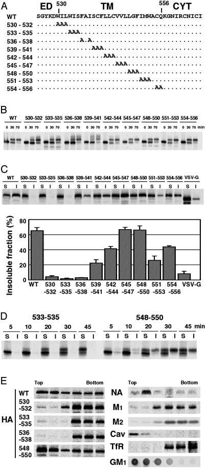
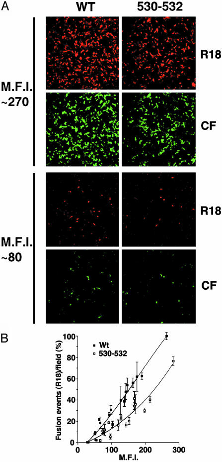
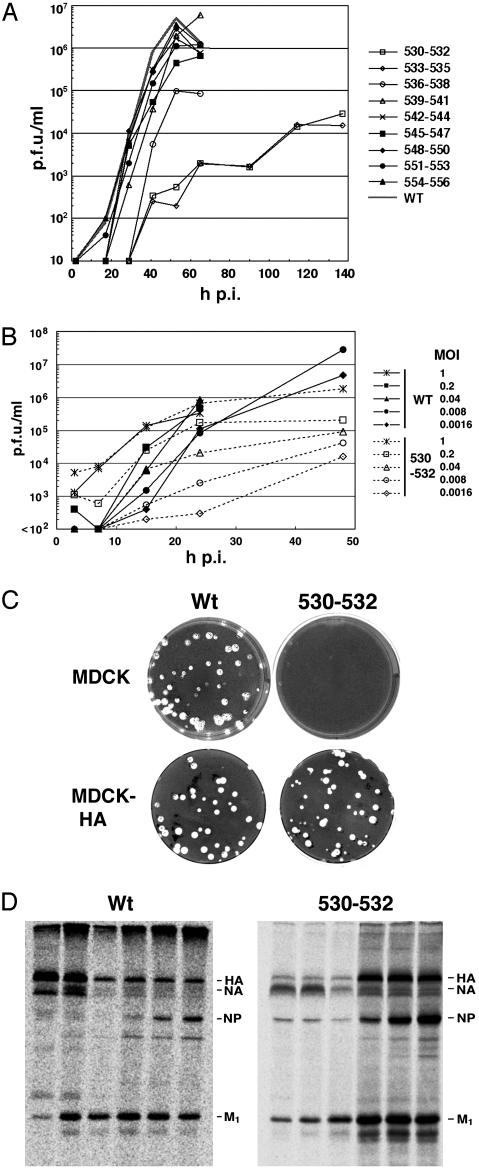
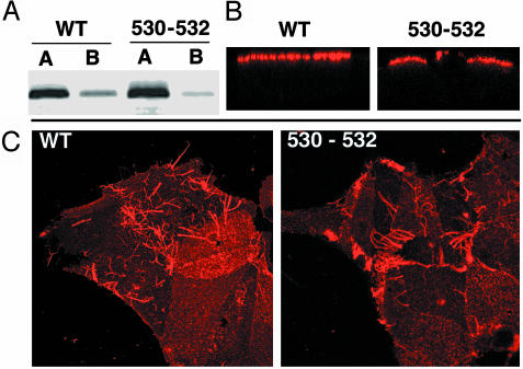
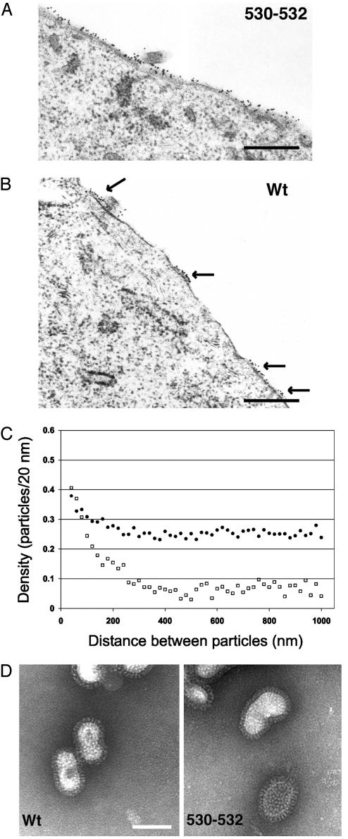
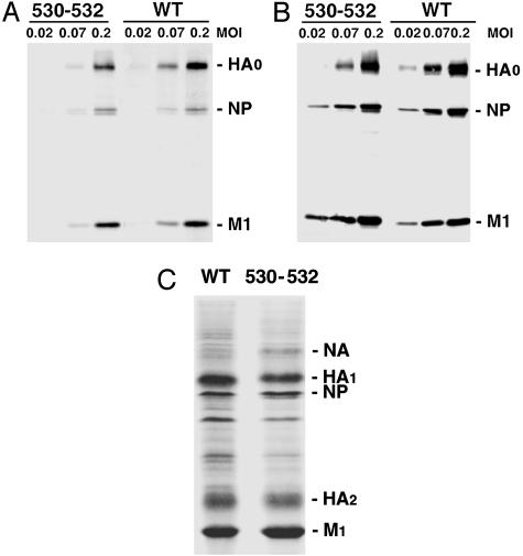
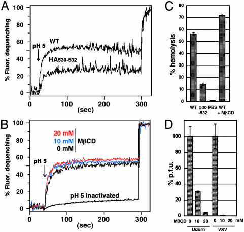
Lipid raft microdomains are enriched in sphingomyelin and cholesterol and function as platforms for signal transduction and as the site of budding of several enveloped viruses, including influenza virus. The influenza virus hemagglutinin (HA) glycoprotein, which mediates both viral-cell attachment and membrane fusion, associates intrinsically with lipid rafts. Residues in the HA transmembrane (TM) domain are important for raft association as sequence substitutions in the HA TM domain ablate HA association with rafts (nonraft HA). Cells expressing either WT or nonraft HA cause complete fusion (lipid mixing and content mixing) over widely varying HA expression levels. However, the number of fusion events measured for nonraft HA mutant protein at all HA surface densities was reduced to approximately 55% of the events for WT HA protein. Mutant influenza viruses were generated that contain the nonraft HA TM domain alterations. Electron microscopy experiments showed that WT HA was distributed at the cell surface in clusters of 200-280 nm in diameter, whereas nonraft HA was distributed mostly randomly at the plasma membrane. Nonraft HA virus showed reduced budding, contained reduced amounts of HA protein, was greatly reduced in infectivity, and exhibited decreased virus-membrane fusion activity. Cholesterol depletion of virus did not affect the ability of virions to cause either virus-cell lipid mixing or virus-mediated hemolysis, a surrogate for content mixing. Taken together, the data suggest that HA clusters in rafts to provide a sufficient concentration of HA in budding virus to mediate efficient virus-cell fusion.


Figures

Fig. 1.
Fig. 1.
Alanine-scanning mutagenesis and characterization of the mutant HA proteins. (A) Schematic diagram showing the amino acid substitutions made in the influenza virus HA TM domain. ED, ectodomain; CYT, cytoplasmic tail. (B) Carbohydrate maturation of WT and mutant HA proteins in MDCK cells stably expressing WT or mutant HAs. (C) Solubility of HA in 0.25% TX-100 at 4°C in BHK cells transiently expressing WT and mutant HAs. S, soluble fractions; I, insoluble fractions. (D) Correlation between HA carbohydrate maturation and TX-100 insolubility in BHK cells transiently expressing HAs. S, soluble fractions; I, insoluble fractions. (E) Flotation sucrose density gradient of 0.25% TX-100-lysed HeLa-T4 cells transiently expressing WT and mutant HAs, NA, M1, and M2 proteins. Cav, caveolin; TfR, transferrin receptor; GM1, GM1 ganglioside. GM1 was detected by a dot blot analysis using a peroxidase-conjugated cholera toxin B subunit.
Fig. 2.
Fig. 2.
Nonraft HA has reduced cell–cell fusion capacity as compared with WT HA at equivalent overall surface density of HA. (A) HA fusion activity of WT and nonraft mutant HA 530–532 was measured by binding RBCs dual-labeled with lipid probe octadecyl rhodamine (R18) and aqueous cytoplasmic probe CF to BHK cells transiently expressing HAs and triggering fusion by low pH treatment (pH 4.8 for 1 min). MFI, mean fluorescent intensity. (B) Quantification of fusion events for BHK cells expressing different cell surface densities of HAs as measured by flow cytometry. Average fusion events and standard deviations are from three to five microscopic fields per experiment. MFI, mean fluorescent intensity.
Fig. 3.
Fig. 3.
Characterization of influenza viruses containing HA TM alanine substitutions: replication kinetics, trans complementation, and flotation gradient analysis. WT influenza virus and nine HA TM domain mutant viruses were recovered by using reverse genetics methods from cloned DNAs. (A) Growth curve of influenza viruses by using a moi of 0.001 pfu per cell. Viral titers were determined by plaque assay on MDCK-HA cells. h p.i., hours postinfection. (B) Effect of different moi on nonraft HA 530–532 virus growth rates. Viruses were grown in MDCK cells at the moi shown and plaqued on MDCK-HA cells. (C) Viral plaques of WT virus and HA 530–532 virus on MDCK and MDCK-HA cells. (D) TX-100 solubilization and flotation analysis of HeLa-T4 cells infected with WT or HA 530–532 viruses.
Fig. 4.
Fig. 4.
Nonraft HA 530–532 is targeted to the apical surface of polarized cells. (A) MDCK cells were grown on 24-mm diameter, 0.4-μm pore size Transwell polycarbonate filters (Costar) and infected with WT or nonraft HA 530–532 virus from the apical cell surface at a moi of 3.0 pfu per cell. Monolayers were biotinylated at 5 h p.i. as described (10) and immunoprecipitated with HA-specific mAb C45/3 (26). Polypeptides were separated by SDS/PAGE and blotted to poly(vinylidene difluoride) membranes, and the surface-biotinylated proteins were detected by using Alexa 680-conjugated streptavidin and binding quantified with an Odyssey Infrared Imaging system (Li-Cor). (B) Localization of HA on polarized virus-infected MDCK cells. Cells were infected as above and stained with mAb C45/3 followed by incubation with Texas red-conjugated donkey anti-goat secondary antibody (Jackson ImmunoResearch). Fluorescence was observed with a Zeiss LSM 410 confocal microscope. Z-scans are shown. (C) Staining of virus-infected MDCK cells with goat anti-Udorn serum to examine whether production of filamentous virus production (a characteristic of field-isolated influenza A viruses including the A/Udorn/72 strain) depends on the interaction with rafts.
Fig. 5.
Fig. 5.
Distribution of HA at the plasma membrane: WT HA but not nonraft HA is clustered in microdomains. (A and B) MDCK cells were infected with nonraft HA virus (A) and WT virus (B) at a moi of 5 pfu per cell, and at 4.5 h p.i. cells were incubated with mAb C78/1 specific for Udorn HA protein followed by a secondary antibody conjugated to 15-nm gold particles and processed for sectioning, staining, and electron microscopy. Cell profiles were randomly selected and photographed at ×36,000. (Bar = 0.5 μm.) (C) The plasma membrane length and the gold particle distribution of ≈30 random cell profiles were measured by using the method of Brown and Lyles (28). •, Nonraft HA virus. □, WT virus. (D) Negatively stained purified influenza virions. Images are at ×89,360. (Bar = 100 nm.)
Fig. 6.
Fig. 6.
Nonraft HA virions have decreased HA protein composition. (A and B) Protein composition of WT and nonraft HA virus. MDCK cells were infected with WT and nonraft mutant (530–532) viruses at varying moi indicated. At 18 h p.i., the culture media (A) and the cells (B) were harvested. The polypeptide composition of both cell lysates and purified virions was analyzed by immunoblotting using an anti-influenza virus specific serum. The ratio of HA, NP, and M1 proteins in virions as compared with cells was quantified. (C) Coomassie brilliant blue-stained polypeptides of purified WT and mutant (530–532) viruses grown in MDCK cells are shown.
Fig. 7.
Fig. 7.
Nonraft HA virus has reduced fusion activity and does not require cholesterol for fusion. (A) Virus–RBC ghost fusion was assayed by fluorescence dequenching. Purified WT and nonraft 530–532 virions were labeled with R18 and bound to sealed human RBC ghosts. Fusion between the R18-labeled virus and the RBC ghosts was triggered by lowering the pH from 7.0 to 5.0 (arrow). After 300 s of fluorescence recording TX-100 was added to the cuvette to obtain the maximum dequenching. (B) Fusion activity of MβCD-treated virus. R18-labeled WT virus was incubated in PBS (pH 7.0) for 15 min at 37°C in the absence or presence of 10 mM or 20 mM MβCD, and virions were repurified through a sucrose cushion. Fusion kinetics between the viruses and ghosts were analyzed as described above. To inactivate HA protein the R18-labeled virus was incubated in PBS (pH 5.0) for 15 min at 37°C. (C) Hemolysis assay as a surrogate for membrane fusion content mixing. WT and nonraft 530–532 virus (3.3 μg protein) or PBS were incubated with human RBCs for 45 min at 4°C. The RBCs were then incubated in a low pH buffer (PBS pH 5.0) for 1 min at room temperature. After replacing the low pH buffer with a neutral pH PBS, the RBCs were incubated for 5 min at 37°C. The amount of released hemoglobin was determined spectrophotometrically at 540 nm. The hemolysis capacity of WT virus treated with 20 mM MβCD (WT + MβCD) was also determined. RBCs lysed in 1% TX-100 was set at 100% hemolysis. (D) Reduction of plaque-forming capacity by MβCD. Influenza virus and VSV were incubated in PBS (pH 7.0) for 15 min at 37°C in the absence or presence of 10 mM or 20 mM MβCD. MβCD was removed by pelleting the virus through a 30% sucrose cushion, and infectivity of the pelleted virus was determined by plaque assay. Pfu of the MβCD-untreated viruses are shown as 100%.

References

    1. Simons, K. & Ikonen, E. (1997) Nature 387, 569–572. - PubMed
    1. Suomalainen, M. (2002) Traffic 3, 705–709. - PubMed
    1. Pralle, A., Keller, P., Florin, E.-L., Simons, K. & Horber, J. (2000) J. Cell Biol. 148, 997–1007. - PMC - PubMed
    1. Varma, R. & Mayor, S. (1998) Nature 394, 798–801. - PubMed
    1. Simons, K. & Toomre, D. (2000) Nat. Rev. Mol. Cell. Biol. 1, 31–39. - PubMed

Publication types

MeSH terms

LinkOut - more resources