Comparative Study
. 2003 Oct 20;89(8):1463-72.
doi: 10.1038/sj.bjc.6601243.

Altered maturation of peripheral blood dendritic cells in patients with breast cancer

Affiliations
Comparative Study

Altered maturation of peripheral blood dendritic cells in patients with breast cancer

S Della Bella et al. Br J Cancer. .

Abstract

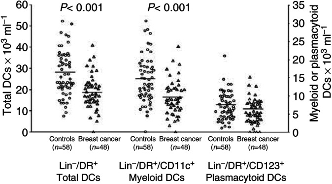
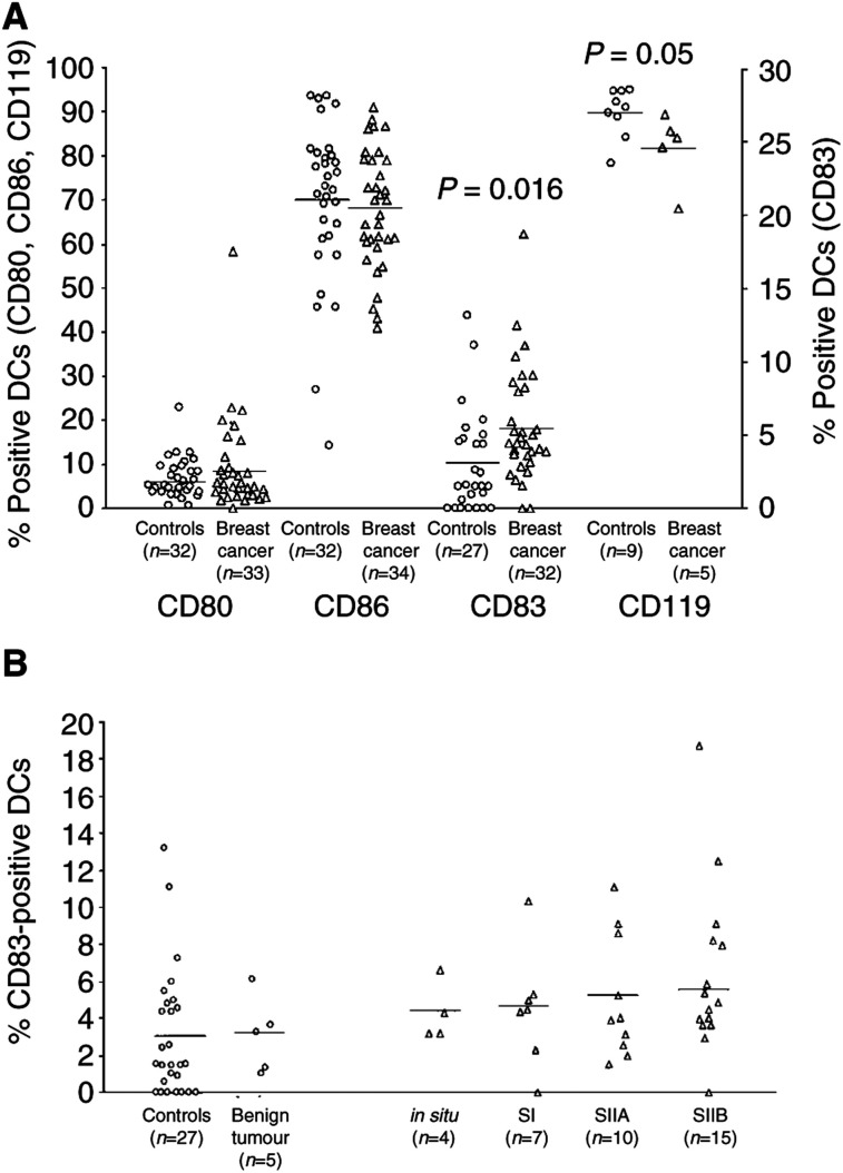
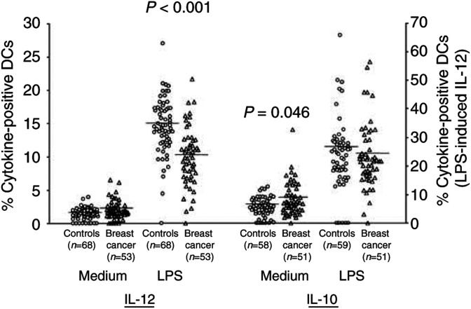
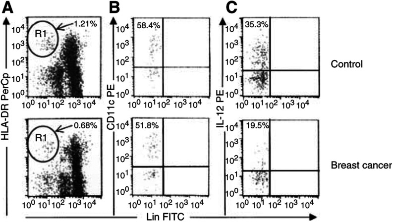
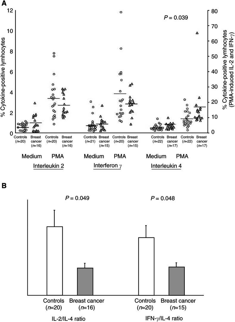
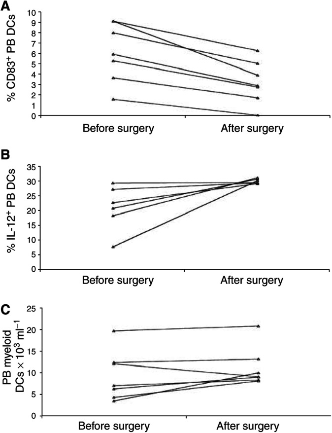
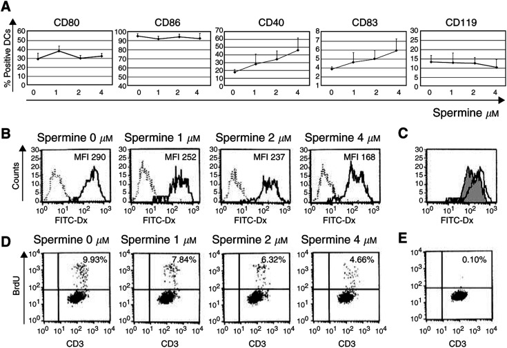
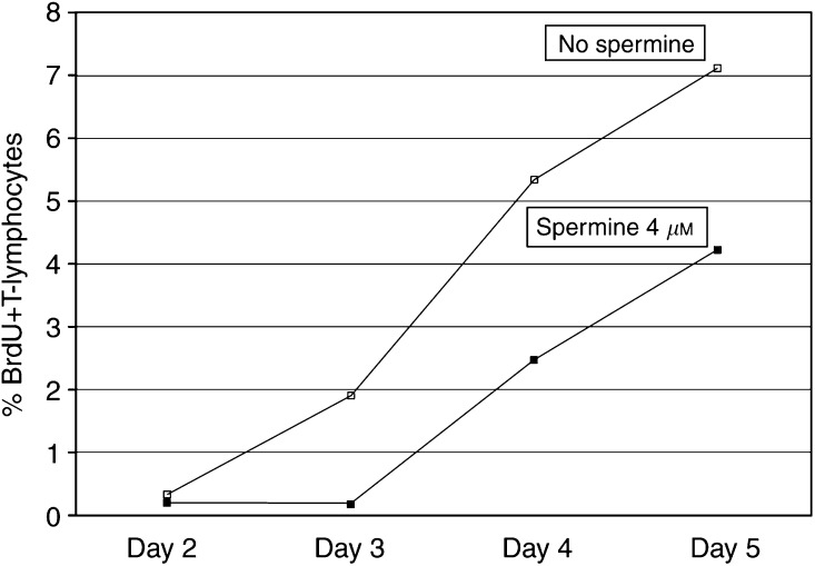
Tumours have at least two mechanisms that can alter dendritic cell (DC) maturation and function. The first affects the ability of haematopoietic progenitors to differentiate into functional DCs; the second affects their differentiation from CD14+ monocytes, promoting an early but dysfunctional maturation. The aim of this study was to evaluate the in vivo relevance of these pathways in breast cancer patients. For this purpose, 53 patients with invasive breast cancer were compared to 68 healthy controls. To avoid isolation or culture procedures for enrichment of DCs, analyses were directly performed by flow cytometry on whole-blood samples. The expression of surface antigens and intracellular accumulation of regulatory cytokines upon LPS stimulation were evaluated. The number of DCs, and in particular of the myeloid subpopulation, was markedly reduced in cancer patients (P<0.001). Patient DCs were characterized by a more mature phenotype compared with controls (P=0.016), and had impaired production of IL-12 (P<0.001). These alterations were reverted by surgical resection of the tumour. To investigate the possible role of some tumour-related immunoactive soluble factors, we measured the plasmatic levels of vascular endothelial growth factor, IL-10 and spermine. A significant inverse correlation between spermine concentration and the percentage of DCs expressing IL-12 was found. Evidence was also obtained that in vitro exposure of monocyte-derived DCs to spermine promoted their activation and maturation, and impaired their function. Taken together, our results suggest that both the above-described mechanisms could concomitantly act in breast cancer to affect DC differentiation, and that spermine could be a mediator of dysfunctional maturation of DCs.


Figures

Figure 1
Figure 1
Absolute number of PB DCs in breast cancer patients compared with controls. A significant decrease in PB Lin-/HLA-DR+ cells (left axis) and myeloid DCs (right axis) was observed in cancer patients. Whole blood was stained with a cocktail of FITC-conjugated mAbs recognising CD3, CD14, CD16, CD19 and CD20, and with PerCP-conjugated anti-HLA-DR mAb; myeloid DCs were identified as lineage-/HLA-DR+/CD11c+ cells, plasmacytoid DCs (right axis) as lineage-/HLA-DR+/CD123+ cells. Absolute DC counts were then determined indirectly by multiplying the percentage of DCs in the mononuclear gate times the sum of the lymphocyte and monocyte determined on a differential blood cell counter. Each symbol represents a single sample. Open circles: control subjects; open triangles: breast cancer patients. Mean values represented by horizontal lines in each series. P-values were determined using the t-test for independent samples, patients compared with controls.
Figure 2
Figure 2
Immunophenotype of PB DCs. (A) The activation state of Lin-/HLA-DR+ DCs, assessed as the expression of the costimulatory molecules CD80 and CD86 (left axis), was similar in patients and controls, while the frequency of mature DCs was significantly higher in breast cancer individuals, as assessed by higher percentages of Lin-/HLA-DR+ cells expressing CD83 (right axis) and lower percentages of Lin-/HLA-DR+ cells expressing CD119 (left axis). (B) The percentage of mature DCs, identified as Lin-/HLA-DR+ cells expressing the CD83 maturation marker, increased progressively with the severity of breast cancer. Each symbol represents a single sample. Open circles: control subjects; open triangles: breast cancer patients. Mean values are represented by horizontal lines in each series. P-values were determined using the t-test for independent samples, patients compared with controls.
Figure 3
Figure 3
Cytokine production by peripheral blood DCs in breast cancer patients compared with control subjects. A significantly lower percentage of PB DCs producing IL-12 upon LPS stimulation was observed in cancer patients. After incubation for 5 h in the presence or absence of LPS with BFA added during the last 4 h, whole-blood samples were stained with a cocktail of FITC-conjugated mAbs recognising CD3, CD14, CD16, CD19 and CD20, and with PerCP-conjugated anti-HLA-DR mAb. Intracellular accumulation of cytokines within DCs, identified as lineage-/HLA-DR+ cells, was evaluated after staining with PE-conjugated mAbs directed against either IL-12 or IL-10. Each symbol represents a single sample. Open circles: control subjects; open triangles: breast cancer patients. LPS-induced IL-12 values referred to the right axis; all the other values to the left axis. Mean values represented by horizontal lines in each series. P-values were determined using the t-test for independent samples, patients compared with controls.
Figure 4
Figure 4
Identification and characterisation of PB DCs in whole peripheral blood samples. Comparison between representative flow cytometric analyses from a control subject (upper line) and a breast cancer patient (lower line). (A) Gated on a mononuclear cell analysis region, DCs were identified on the basis of their lack of labelling for the lineage markers CD3, CD14, CD16, CD19 and CD20, but positive staining for HLA-DR (R1). (B) Gated on R1 events, myeloid DCs were identified for their surface expression of CD11c. (C) Gated on R1 events, PB DCs producing IL-12 upon LPS stimulation were identified for their intracellular accumulation of the cytokine.
Figure 5
Figure 5
Cytokine production by T lymphocytes in breast cancer patients compared with control subjects. PBMCs were cultured in the presence of BFA with or without PMA plus ionomycin for 18 h. At the end of the cultures, cells were stained with FITC-conjugated anti-CD3 mAb, and intracellular accumulation of cytokines within CD3+ T lymphocytes was evaluated after staining with PE-conjugated mAbs directed against either IL-2 or IFN-γ or IL-4. (A) Each symbol represents a single sample. Open circles: control subjects; open triangles: breast cancer patients. Values of PMA-induced IL-2 and IFN-γ referred to the right axis, all the others to the left axis. Mean values are represented by horizontal lines in each series. P-values were determined using the t-test for independent samples, patients compared with controls. (B) Profile of cytokine production by T lymphocytes, expressed as type 1 to type 2 cytokine ratios. Both IL-2 to IL-4 and IFN-γ to IL-4 ratios were significantly reduced in breast cancer patients compared with controls. Open bars: control subjects; dark bars: breast cancer patients. P-values were determined using the t-test for independent samples, patients compared with controls.
Figure 6
Figure 6
Effects of surgical removal of the tumour on PB DCs. At 4 weeks after surgery, (A) the percentage of CD83+ PB DCs significantly decreased (P=0.001), with a reduction of mature PB DCs observed in all the patients; (B) the percentage of PB DCs expressing IL-12 upon LPS stimulation significantly increased (P=0.042), with a complete normalisation observed in all the patients; (C) the number of myeloid DCs in the peripheral blood of the patients only slightly increased (P=n.s.). Methods as described in Figures 2–4 Each symbol represents a single sample. P-values were determined using the t-test for paired samples.
Figure 7
Figure 7
Effects of spermine on in vitro cultured DCs. Monocyte-derived DCs were generated from the adherent fraction of PBMCs obtained from healthy control subjects in the presence of IL-4 and GM-CSF for 5 days. Spermine at the indicated concentrations was added at the initiation of culture and maintained throughout. (A) Exposure of maturing DCs to spermine affected their immunophenotype, promoting a dose-dependent mild increase of DC expression of CD83 and CD40. Data shown are mean ± s.e.m of four independent experiments. (B) The addition of spermine to maturing DCs impaired their mannose-receptor-mediated FITC-dextran endocytosis, as assessed by a dose-dependent mean fluorescence intensity reduction. Dashed lines indicate FITC-dextran endocytosis at 0°C (negative control), and solid lines represent FITC-dextran endocytosis at 37°C at the indicated spermine concentrations. This result is representative of four independent experiments. (C) Same experiment as (B). Comparison between FITC-dextran endocytosis by DCs cultured in the absence (white histogram) or presence (grey histogram) of 4 μM spermine. (D) Exposure of maturing DCs to spermine affected their allostimulatory capacity. Lymphocyte proliferation was measured by flow cytometry as BrdU incorporation by CD3+ cells. The percentages of proliferating allogenic T lymphocytes after coculture for 5 days with DCs generated in the presence of the indicated spermine concentrations are presented. This result is representative of four independent experiments. (E) BrdU incorporation by T lymphocytes cultured for 5 days in the absence of DCs (negative control) is shown.
Figure 8
Figure 8
Analysis of T-cell division induced at different days by allogenic unconditioned (white squares) or spermine-conditioned (black squares) DCs. The decrease in T-cell stimulatory function seen for DCs cultured with spermine was not due to a different cell kinetics. Methods as in Figure 7; results expressed as percentages of proliferating allogenic T lymphocytes.

Similar articles

Cited by

References

    1. Almand B, Resser JR, Lindman B, Nadaf S, Clark JI, Kwon ED, Carbone DP, Gabrilovich DI (2000) Clinical significance of defective dendritic cell differentiation in cancer. Clin Cancer Res 6: 1755–1766 - PubMed
    1. Almeida J, Bueno C, Alguero MC, Sanchez ML, Canizo MC, Fernandez ME, Vaquero JM, Laso FJ, Escribano L, San Miguel JF, Orfao A (1999) Extensive characterization of the immunophenotype and pattern of cytokine production by distinct subpopulations of normal human peripheral blood MHC+/lineage- cells. Clin Exp Immunol 118: 392–401 - PMC - PubMed
    1. Arpinati M, Green CL, Heimfeld S, Heuser JE, Anasetti C (2000) Granulocyte-colony stimulating factor mobilizes T helper 2-inducing dendritic cells. Blood 95: 2484–2490 - PubMed
    1. Bell D, Chomarat P, Broyles D, Netto G, Harb GM, Lebecque S, Valladeau J, Davoust J, Palucka KA, Banchereau J (1999) In breast carcinoma tissue, immature dendritic cells reside within the tumour, whereas mature dendritic cells are located in peritumoral areas. J Exp Med 190: 1417–1425 - PMC - PubMed
    1. Bellone M, Iezzi G, Rovere P, Galati G, Ronchetti A, Protti MP, Davoust J, Rugarli C, Manfredi AA (1997) Processing of engulfed apoptotic bodies yields T cell epitopes. J Immunol 159: 5391–5399 - PubMed

Publication types