. 2004 Feb 15;103(4):1475-84.
doi: 10.1182/blood-2003-06-2116. Epub 2003 Oct 16.

E mu-BRD2 transgenic mice develop B-cell lymphoma and leukemia

Affiliations

E mu-BRD2 transgenic mice develop B-cell lymphoma and leukemia

Rebecca J Greenwald et al. Blood. .

Abstract

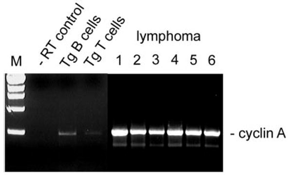
Transgenic mice with lymphoid-restricted overexpression of the double bromodomain protein bromodomain-containing 2 (Brd2) develop splenic B-cell lymphoma and, upon transplantation, B-cell leukemia with leukemic infiltrates in liver and lung. Brd2 is a nuclear-localized transcription factor kinase that is most closely related to TATA box binding protein-associated factor, 250 kDa (TAF(II)250) and the Drosophila developmental protein female sterile homeotic. Constitutive expression of BRD2 in the lymphoid compartment increases cyclin A transcription, "priming" transgenic B cells for proliferation. Mice stochastically develop an aggressive B-cell lymphoma with the features of B-1 cells, including CD5 and surface IgM expression. The B-cell lymphoma is monoclonal for immunoglobulin gene rearrangement and is phenotypically stable. The lymphoblasts are very large and express a transcriptome that is similar to human non-Hodgkin lymphomas. Both a wild-type BRD2 transgene and a kinase-null point mutant drive lymphomagenesis; therefore we propose that, rather than kinase activity, Brd2-mediated recruitment of E2 promoter binding factors (E2Fs) and a specific histone acetyltransferase to the cyclin A promoter by both types of transgene is a mechanistic basis for neoplasia. This report is the first to describe a transgenic mouse model for constitutive expression of a protein with more than one bromodomain.


Figures

Figure 1
Figure 1. Expression of transgene in lymphoid tissues
(A) RT-PCR analysis of Eμ-BRD2 expression in different Tg tissues. Lane 1, non-Tg spleen control; lane 2, Tg heart control; lane 3, Tg skeletal muscle control; lane 4, Tg spleen; lane 5, Tg thymus. Primers amplified both endogenous murine message and Tg message. Negative controls without RT verified the RT dependence of the signal (results not shown). (B) Tg expression in Tg B and T cells. The 5′ primer recognizes a Tg vector–specific sequence at the translation start site and the 3′ primer a sequence within the first bromodomain, common to both endogenous and Tg sequences. Analysis was performed with purified B-cell RNA (B220) and T-cell RNA (CD3). Control reaction without reverse transcriptase is shown (−RT control).
Figure 2
Figure 2. Proliferation of Tg and Tgmut B cells in response to anti-CD40 and anti-IgM stimulation
B cells from spleens of littermate control (■), Tg line 28 (formula image
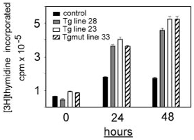
), Tg line 23 (□), or Tgmut line 33 (▨) or were stimulated in vitro for 24 or 48 hours with anti-CD40 and anti-IgM. Incorporation of [3H] thymidine is shown with standard deviation (n = 4) for each bar.
Figure 3
Figure 3. Flow cytometry of lymphoma cells
(A) Unfractionated splenocytes from mice diagnosed with splenic lymphoma and peripheral leukemia (lymphoma) were stained for various markers of B-cell differentiation and activation and compared with asymptomatic non-Tg littermate splenocytes (control). Numbers in each square refer to percentages of gated events in a particular quadrant. (B) Peritoneal lymphocytes of premalignant, asymptomatic Tg mice (asymptomatic) were compared with age-matched littermate (control) to assess expansion of the B-1 cell population in the peritoneum.
Figure 4
Figure 4. Tg-driven splenic lymphoma and peripheral leukemia
(A) A B220-stained section of asymptomatic Tg spleen (i; bar, 200 μm) is compared with a B220-stained section of a Tg mouse (ii; bar, 100 μm) or Tgmut mouse (iii; bar, 100 μm) with lymphocytic lymphoma. An H&E-stained section of asymptomatic Tg spleen (iv; bar, 50 μm) is compared with an H&E-stained section of a Tg mouse with lymphocytic lymphoma (v; bar, 50 μm). An H&E-stained section of bone marrow of Tgmut mouse with lymphocytic lymphoma is also shown (vi; bar, 100 μm). (B) A Wright-Giemsa–stained smear of peripheral blood of a leukemic mouse that received Tg B-cell lymphoma transplants shows overproliferation of lymphoblasts (i; bar, 50 μm). Blasts exhibit prominent, multiple chromocenters and scanty dark blue cytoplasm (ii; bar, 20 μm). Note the polymorphonuclear granulocyte for comparison. Additional field emphasizes homotypic adhesion and mixed chromatin states (iii; bar, 20 μm).
Figure 5
Figure 5. Immunoglobulin gene rearrangements are monoclonal
(A) Ig RNAs were isolated from purified B cells of a Tg splenic B-cell lymphoma, amplified by RT-PCR, and resolved. −RT control is shown (left). Primers to the 3′ constant region (3′Cμ and 3′Cκ) Ig sequences and degenerate primers to the variable regions (5′VH and Vk) were used in pairs as indicated. RNA of purified, polyclonal B cells from normal, non-Tg littermate control (right) is compared with Tg lymphoma (left). (B) Lymphoma RNA from first or sixth transplanted passage of malignancy was amplified with primers specific for Bcl6 (■) or Blimp-1 genes (□) by real-time PCR and compared with splenic B2 control. Relative expression levels were determined by the 2−ΔΔCt method where ΔΔCt = ΔCt, sample − ΔCt, reference with Bcl6 or Blimp-1 serving as samples and β2-microglobulin as a reference.
Figure 6
Figure 6. Kaplan-Meier plots showing survival of mice that received Tg B-cell lymphoma transplants
Non-Tg mice were irradiated with 6 Gy (dashed line and dotted line) or 8 Gy (solid line) and inoculated subcutaneously with 107 purified B cells from the spleen of a Tg donor with lymphoma (dotted line and solid line) or 107 purified B cells from the spleen of an asymptomatic Tg donor (dashed line). For dashed line, n = 4; for dotted line, n = 6; and for solid line, n = 15. Control indicates B cells from asymptomatic Tg donor; and DLCL, diffuse large B-cell lymphoma.
Figure 7
Figure 7. Cyclin A expression in Tg B-cell malignancy
RT-PCR was performed with RNA from purified B cells (Tg B cells) or purified T cells (Tg T cells) from spleens of asymptomatic Tg mice, using primers to the cyclin A gene. Results were compared with negative control (−RT) and to RNA from B cells obtained from independently arising B-cell lymphomas in 6 different mice. Lymphoma lane 1, Tg leukemic mouse (transplant donor); lane 2, Tgmut leukemic mouse; lane 3, second serial transplantation; lane 4, second serial transplantation; lane 5, fourth serial transplantation; lane 6, fourth serial transplantation.

References

    1. Denis GV. Bromodomain motifs and “scaffolding”? Front Biosci. 2001;6:D1065–D1068. - PMC - PubMed
    1. Horn PJ, Peterson CL. The bromodomain: a regulator of ATP-dependent chromatin remodeling? Front Biosci. 2001;6:D1019–1023. - PubMed
    1. Zeng L, Zhou MM. Bromodomain: an acetyl-lysine binding domain. FEBS Lett. 2002;513:124–128. - PubMed
    1. Haynes SR, Dollard C, Winston F, Beck S, Trowsdale J, Dawid IB. The bromodomain: a conserved sequence found in human, Drosophila and yeast proteins. Nucleic Acids Res. 1992;20:2603. - PMC - PubMed
    1. Jeanmougin F, Wurtz JM, Le Douarin B, Chambon P, Losson R. The bromodomain revisited. Trends Biochem Sci. 1997;22:151–153. - PubMed

Publication types

MeSH terms