. 2003 Nov 1;17(21):2675-87.
doi: 10.1101/gad.275203. Epub 2003 Oct 16.

Meiotic double-strand breaks at the interface of chromosome movement, chromosome remodeling, and reductional division

Affiliations

Meiotic double-strand breaks at the interface of chromosome movement, chromosome remodeling, and reductional division

Aurora Storlazzi et al. Genes Dev. .

Abstract

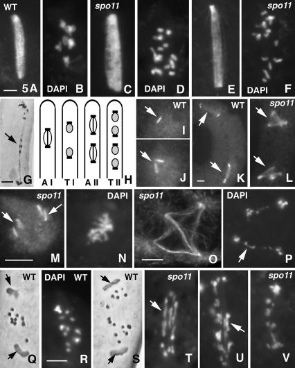
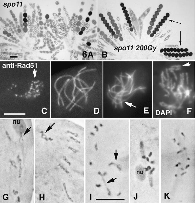
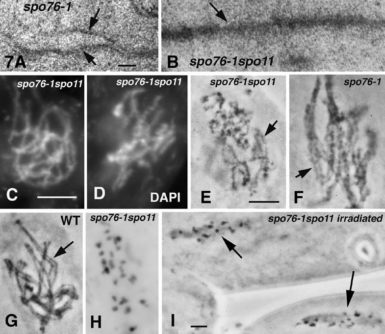
Chromosomal processes related to formation and function of meiotic chiasmata have been analyzed in Sordaria macrospora. Double-strand breaks (DSBs), programmed or gamma-rays-induced, are found to promote four major events beyond recombination and accompanying synaptonemal complex formation: (1) juxtaposition of homologs from long-distance interactions to close presynaptic coalignment at midleptotene; (2) structural destabilization of chromosomes at leptotene/zygotene, including sister axis separation and fracturing, as revealed in a mutant altered in the conserved, axis-associated cohesin-related protein Spo76/Pds5p; (3) exit from the bouquet stage, with accompanying global chromosome movements, at zygotene/pachytene (bouquet stage exit is further found to be a cell-wide regulatory transition and DSB transesterase Spo11p is suggested to have a new noncatalytic role in this transition); (4) normal occurrence of both meiotic divisions, including normal sister separation. Functional interactions between DSBs and the spo76-1 mutation suggest that Spo76/Pds5p opposes local destabilization of axes at developing chiasma sites and raise the possibility of a regulatory mechanism that directly monitors the presence of chiasmata at metaphase I. Local chromosome remodeling at DSB sites appears to trigger an entire cascade of chromosome movements, morphogenetic changes, and regulatory effects that are superimposed upon a foundation of DSB-independent processes.


Figures

Figure 1.
Figure 1.
Sordaria Spo11p. Indicated are the five conserved regions (rectangles with different motifs), intron location, nuclear localization signal (NLS), and sites of 11 spo11 mutations. (black triangles) Predicted stop codons; (flag) tyrosine spo11-Y171F mutation; (Δ) deletion.
Figure 2.
Figure 2.
Spo11p localization. Spo11-GFP staining (left) and corresponding DAPI (right). Karyogamy nucleus (A,B) and early leptotene nucleus (C,D; double-stained with anti-GFP antibody) showing chromatin-associated Spo11-GFP foci (arrows). (E,F) Mid-leptotene nucleus (anti-GFP antibody); foci are mainly in rows (arrows). (G,H) Leptotene nucleus with clear presynaptic alignment and Spo11-GFP foci in rows (arrow). (I,J) Spo11-GFP stains as discontinuous narrow lines along all chromosomes of late leptotene and bouquet nuclei (arrow). (K,L) Late pachytene nucleus with diffuse Spo11p signal. (M,N) Ascospore with two nuclei resulting from the second postmeiotic division stained by Spo11-GFP and DAPI. Bars, 5 µm.
Figure 3.
Figure 3.
Prophase phenotypes of wild type and spo11 mutants. Nuclei are stained by DAPI (A,B,D,E) and by hematoxylin (C,F). (A-C) Wild-type. (A) Leptotene. (B) Diffuse stage. (C) Diplotene nuclei. (D-F) Corresponding spo11Δ nuclei. (F) Note similar chromosome condensation at diplotene despite no chiasmata among the 14 univalents; arrows point to the two widely separated homologs attached to the nucleolus (nu). (G-J) Chromosome axes are visualized by Spo76-GFP. (G,H) Early and mid-prophase nuclei of spo11Δ. Note partial alignment of one pair of homologs (arrow in H). (I,J) Wild-type nuclei at equivalent ascus sizes. (I) Zygotene; arrow points to aligned segments at the same distance as aligned segment noted in H. (J) Pachytene with seven synapsed bivalents. (K-M) Electron micrographs of thin sections of AEs from wild-type (K) and spo11-1 (L,M) nuclei. Note striations (arrows) in wild-type and mutant AEs. (M) Late prophase spo11 AE shows thicker, separated sister axes. (N) Three-dimensional representation of a reconstructed spo11-1 nucleus (from 56 sections/pictures); arrows indicate an aligned homolog pair. Bars: LM pictures, 5 µm; EM pictures, 0.1 µm.
Figure 4.
Figure 4.
Wild-type and spo11 bouquet. Chromosome axes are stained by Spo76-GFP. Wild-type late leptotene (A; note aligned homologs) and early pachytene nucleus (B) with respectively loose and tight bouquets. spo11Δ early (C) and later (D) bouquet. Telomere clustering releases at late prophase in both spo11-Y171F (E) and wild type (F; note the seven synapsed bivalents). Bar, 5 µm.
Figure 5.
Figure 5.
Division I and II in wild-type and spo11 asci. Spindles and SPBs are stained with anti-α-tubulin and anti-MPM2 antibodies, respectively. Wild-type MI spindle (A) and corresponding seven bivalents (B) stained by DAPI. (C-G) spo11Δ nuclei. MI spindle (C) and corresponding DAPI showing 14 univalents (D). (E,F) AI spindle and DAPI. (G) Late AI (hematoxylin) showing laggard chromosomes (arrow). (H) Drawing of wild-type asci at anaphase I (AI), telophase I (TI), anaphase II (AII), and telophase II (TII). (I) One of the two wild-type TI nuclei with one SPB (arrow). (J) Wild-type prophase II nucleus showing duplicated SPBs (arrow). (K) Wild-type ascus with two division II bipolar spindles (arrows) and four SPBs. (L) Corresponding spo11Δ ascus with two tetrapolar division II spindles (arrows). (M) One of the two spo11Δ prophase II nuclei with four SPBs (arrows; cf. J). (N) Corresponding DAPI. (O) Tetrapolar spo11Δ AII spindle with clear astral microtubules emanating from the four poles and crossed intranuclear microtubules. (P) Corresponding DAPI: Both segregating sets of chromosomes show laggards (arrow). (Q) Wild-type MII stained with hematoxylin: Three chromosomes show separating chromatids. Arrows point to the two SPBs. (R) Wild-type early AII stained by DAPI; note that hematoxylin and DAPI give similar chromosome pictures. (S) Wild-type AII (hematoxylin); 14 sister chromatids separate and segregate regularly (arrows point to SPBs). (T) In contrast, spo11 MII show elongated chromosomes (arrow). (U,V) The two spo11 AII nuclei from one ascus; both show stretched chromosomes (arrow in U) and a mixture of chromosomes and chromatids (smaller units). Bars, 5 µm.
Figure 6.
Figure 6.
Phenotypes of irradiated spo11 mutants. (A) spo11Δ asci (from one perithecium) form only few ascospores. (B) After γ irradiation, eight-spored asci are regularly formed (arrows). (C-J) γ-Irradiated spo11Δ asci. (C) Leptotene nucleus with numerous Rad51 foci (arrow); anti-Rad51 antibody staining. (D,E) Spo76-GFP staining. (D) Restored pachytene nucleus with seven bivalents (cf. Fig. 3H). (E) Nucleus with only partial synapsis (arrow). (F) DAPI-stained nucleus showing broken chromosomes (arrow; cf. their sizes and chromosomes in D). (G-K) Chromosomes are stained by hematoxylin. (G-I) Restored diplotene nuclei with clear chiasmata. (G) All bivalents exhibit at least one chiasma. (H) The seven bivalents show several chiasmata each. Arrows point to the nucleolar organizer bivalent 2 (nu), exhibiting two (G) and four (H) chiasmata. (I) Nucleus with only two chiasmata (arrows). (J) MI spindle with seven bivalents; the nucleolar bivalent (left) is still attached to the nucleolus (nu). (K) Control unirradiated spo11Δ MI nucleus with 14 univalents. Bars, 5 µm.
Figure 7.
Figure 7.
spo11-1 spo76-1 phenotypes. (A) Electron micrograph of a spo76-1 AE; note clear progressive AE splitting (arrows) from right to left. (B) spo76-1 spo11 longitudinally sectioned AE (arrow). (C,D) Early and late prophase nuclei of spo11-1 spo76-1 stained by DAPI: Chromosomes are more kinky and diffuse at later stages (D) than at early leptotene (cf. chromosomes of single spo11 mutants, Fig. 3D). (E-I) Hematoxylin staining. spo11 spo76-1 chromosomes are unpaired (E) and show similar kinkiness (arrow) as paired spo76-1 chromosomes (F; arrow). (G) Wild-type paired chromosomes are smooth and straight (arrow). (H) spo11 spo76-1 onset of AI with 28 chromatids. (I) Two asci of irradiated spo11 spo76-1: All nuclei arrest at AI. Bars: LM picture, 5 µm; EM pictures, 0.1 µm.

References

    1. Bailis J.M. and Roeder, G.S. 1998. Synaptonemal complex morphogenesis and sister-chromatid cohesion require Mek1-dependent phosphorylation of a meiotic chromosomal protein. Genes & Dev. 12: 3551-3563. - PMC - PubMed
    1. Baum P., Yip, C., Goetsch, L., and Byers, B. 1988. A yeast gene essential for regulation of spindle pole duplication. Mol. Cell Biol. 8: 5386-5397. - PMC - PubMed
    1. Bergerat A., de Massy, B., Gadelle, D., Varoutas, P.C., Nicolas, A., and Forterre, P. 1997. An atypical topoisomerase II from Archaea with implications for meiotic recombination. Nature 386: 414-417. - PubMed
    1. Bishop D.K. 1994. RecA homologs Dmc1 and Rad51 interact to form multiple nuclear complexes prior to meiotic chromosome synapsis. Cell 79: 1081-1092. - PubMed
    1. Blat U., Protacio, R.U., Hunter, N., and Kleckner, N. 2002. Physical and functional interactions among basic chromosome organizational features govern early steps of meiotic chiasma formation. Cell 111: 1-12. - PubMed

Publication types

LinkOut - more resources