Comparative Study
. 2003 Nov;185(21):6255-61.
doi: 10.1128/JB.185.21.6255-6261.2003.

Morphogenesis of bacillus spore surfaces

Affiliations
Comparative Study

Morphogenesis of bacillus spore surfaces

Venkata G R Chada et al. J Bacteriol. 2003 Nov.

Abstract

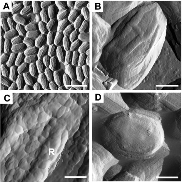
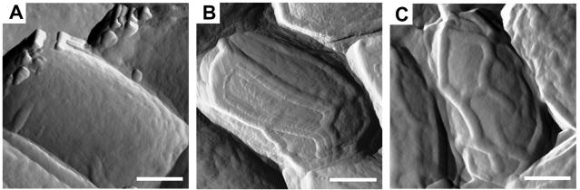
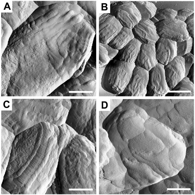
Spores produced by bacilli are encased in a proteinaceous multilayered coat and, in some species (including Bacillus anthracis), further surrounded by a glycoprotein-containing exosporium. To characterize bacillus spore surface morphology and to identify proteins that direct formation of coat surface features, we used atomic-force microscopy (AFM) to image the surfaces of wild-type and mutant spores of Bacillus subtilis, as well as the spore surfaces of Bacillus cereus 569 and the Sterne strain of Bacillus anthracis. This analysis revealed that the coat surfaces in these strains are populated by a series of bumps ranging between 7 and 40 nm in diameter, depending on the species. Furthermore, a series of ridges encircled the spore, most of which were oriented along the long axis of the spore. The structures of these ridges differ sufficiently between species to permit species-specific identification. We propose that ridges are formed early in spore formation, when the spore volume likely decreases, and that when the spore swells during germination the ridges unfold. AFM analysis of a set of B. subtilis coat protein gene mutants revealed three coat proteins with roles in coat surface morphology: CotA, CotB, and CotE. Our data indicate novel roles for CotA and CotB in ridge pattern formation. Taken together, these results are consistent with the view that the coat is not inert. Rather, the coat is a dynamic structure that accommodates changes in spore volume.


Figures

FIG. 1.
FIG. 1.
AFM analysis of B. subtilis spores. Spores were imaged by contact (A) or tapping (B to D) mode, and amplitude image information was collected. The spore in panel D was germinated by exposure to LB medium. Bars: 2.25 μm (A), 375 nm (B), 125 nm (C), 583 nm (D). The “R” in panel C indicates a ridge.
FIG. 2.
FIG. 2.
AFM analysis of B. anthracis and B. cereus spores. Amplitude images of B. anthracis (A and B) or B. cereus (C) spores were collected in tapping mode. Spores with (A) or without (B and C) an exosporium are shown. Bars: 375 nm (A and C), 382 nm (B).
FIG. 3.
FIG. 3.
AFM analysis of B. subtilis mutant spores. Amplitude images of cotE (A), cotA (B), cotB (C) or cotA cotB (D) spores were collected in tapping mode. Bars: 350 nm (A), 2.25 μm (B), 410 nm (C), 468 nm (D).
FIG. 4.
FIG. 4.
Western blot analysis of coat protein cleavage during germination. Proteins were extracted from wild-type spores before (lanes 0) or various times after (indicated in minutes above the lanes) resuspension in LB medium (to initiate germination) or from a cotA mutant (8) (left-most lane in panel B), fractionated by sodium dodecyl sulfate-15% polyacrylamide gel electrophoresis, and subjected to Western blot analysis with anti-CotE (A) or anti-CotA antibodies (B). Arrowheads indicate full-length versions of each protein. Molecular masses are indicated in kilodaltons on either side of the gels.

References

    1. Aronson, A. I., and P. Fitz-James. 1976. Structure and morphogenesis of the bacterial spore coat. Bacteriol. Rev. 40:360-402. - PMC - PubMed
    1. Bauer, T., S. Little, A. G. Stöver, and A. Driks. 1999. Functional regions of the Bacillus subtilis spore coat morphogenetic protein CotE. J. Bacteriol. 181:7043-7051. - PMC - PubMed
    1. Beaman, T. C., H. S. Pankratz, and P. Gerhardt. 1972. Ultrastructure of the exosporium and underlying inclusions in spores of Bacillus megaterium strains. J. Bacteriol. 109:1198-1209. - PMC - PubMed
    1. Catalano, F. A., J. Meador-Parton, D. L. Popham, and A. Driks. 2001. Amino acids in the Bacillus subtilis morphogenetic protein SpoIVA with roles in spore coat and cortex formation. J. Bacteriol. 183:1645-1654. - PMC - PubMed
    1. Charlton, S., A. J. Moir, L. Baillie, and A. Moir. 1999. Characterization of the exosporium of Bacillus cereus. J. Appl. Microbiol. 87:241-245. - PubMed

Publication types

Substances

LinkOut - more resources