. 2003 Nov;185(21):6295-307.
doi: 10.1128/JB.185.21.6295-6307.2003.

Requirement of the Listeria monocytogenes broad-range phospholipase PC-PLC during infection of human epithelial cells

Affiliations

Requirement of the Listeria monocytogenes broad-range phospholipase PC-PLC during infection of human epithelial cells

Angelika Gründling et al. J Bacteriol. 2003 Nov.

Abstract

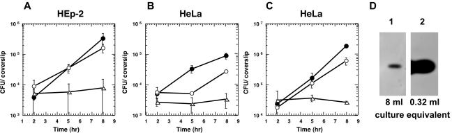
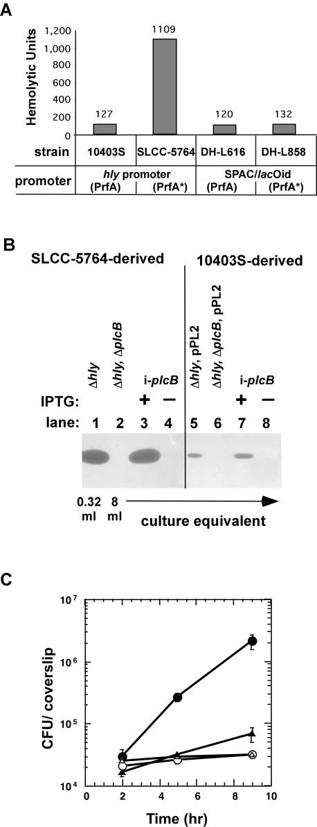
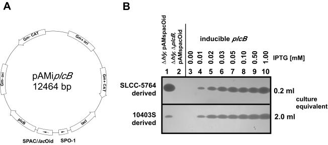
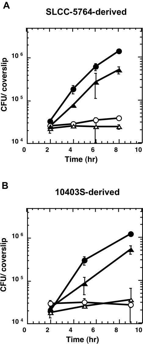
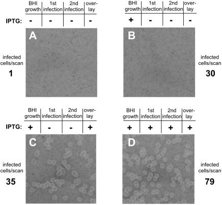
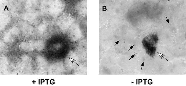
In this study, we investigated the requirement of the Listeria monocytogenes broad-range phospholipase C (PC-PLC) during infection of human epithelial cells. L. monocytogenes is a facultative intracellular bacterial pathogen of humans and a variety of animal species. After entering a host cell, L. monocytogenes is initially surrounded by a membrane-bound vacuole. Bacteria promote their escape from this vacuole, grow within the host cell cytosol, and spread from cell to cell via actin-based motility. Most infection studies with L. monocytogenes have been performed with mouse cells or an in vivo mouse model of infection. In all mouse-derived cells tested, the pore-forming cytolysin listeriolysin O (LLO) is absolutely required for lysis of primary vacuoles formed during host cell entry. However, L. monocytogenes can escape from primary vacuoles in the absence of LLO during infection of human epithelial cell lines Henle 407, HEp-2, and HeLa. Previous studies have shown that the broad-range phospholipase C, PC-PLC, promotes lysis of Henle 407 cell primary vacuoles in the absence of LLO. Here, we have shown that PC-PLC is also required for lysis of HEp-2 and HeLa cell primary vacuoles in the absence of LLO expression. Furthermore, our results indicated that the amount of PC-PLC activity is critical for the efficiency of vacuolar lysis. In an LLO-negative derivative of L. monocytogenes strain 10403S, expression of PC-PLC has to increase before or upon entry into human epithelial cells, compared to expression in broth culture, to allow bacterial escape from primary vacuoles. Using a system for inducible PC-PLC expression in L. monocytogenes, we provide evidence that phospholipase activity can be increased by elevated expression of PC-PLC or Mpl, the enzyme required for proteolytic activation of PC-PLC. Lastly, by using the inducible PC-PLC expression system, we demonstrate that, in the absence of LLO, PC-PLC activity is not only required for lysis of primary vacuoles in human epithelial cells but is also necessary for efficient cell-to-cell spread. We speculate that the additional requirement for PC-PLC activity is for lysis of secondary double-membrane vacuoles formed during cell-to-cell spread.


Figures

FIG. 1.
FIG. 1.
Intracellular growth in human epithelial cells and PC-PLC activity. (A) Monolayers of HEp-2 cells were infected at an MOI of 50:1 with strains 10403S (•), DP-L2161 (10403S Δhly) (○), and DP-L2318 (10403S Δhly ΔplcB) (▵). Intracellular growth was determined as described in Materials and Methods. (B) Monolayers of HeLa cells were infected at an MOI of 67:1 with strains 10403S (•), DP-L2161 (10403S Δhly) (○), and DP-L2318 (10403S Δhly ΔplcB) (▵). (C) Monolayers of HeLa cells were infected at an MOI of 67:1 with PrfA* strains SLCC-5764 (•), DH-L377 (SLCC-5764 Δhly) (○), and DH-L419 (SLCC-5764 Δhly ΔplcB) (▵). The data points in growth curves represent the means ± the standard deviations of three coverslips from one of two experiments. (D) Overnight cultures of strains DP-L2161 (10403S Δhly) and DH-L377 (SLCC-5764 Δhly) were diluted 1:10 in BHI medium and grown for 5 h at 37°C. Proteins from culture supernatants were TCA precipitated and separated by SDS-PAGE. PC-PLC activities were determined by using an egg yolk overlay assay as described in Materials and Methods. Lane 1, the equivalent of 8 ml of DP-L2161 culture supernatant was loaded; lane 2, the equivalent of 0.32 ml of DH-L377 culture supernatant was loaded (1/25 the amount of lane 1).
FIG. 2.
FIG. 2.
Inducible expression of LLO and PC-PLC in L. monocytogenes. (A) Hemolytic activity assay. Hemolytic activities were determined from culture supernatants of L. monocytogenes as described in Materials and Methods. LLO was expressed from the inducible SPAC/lacOid promoter in the presence of 1 mM IPTG or from the native PrfA- or PrfA*-regulated hly promoter in strains 10403S and SLCC-5764, respectively. (B) PC-PLC activity assay. PC-PLC was expressed under the control of the inducible SPAC/lacOid promoter (i-plcB) or the native PrfA-regulated actA-plcB promoter in the L. monocytogenes10403S (PrfA) and SLCC-5764 (PrfA*) backgrounds. Overnight cultures were diluted 1:10 in BHI medium and grown for 5 h at 37°C in the presence or absence of 1 mM IPTG. Culture supernatants were TCA precipitated, and an equivalent of 8 or 0.32 ml of culture supernatant was separated by SDS-PAGE. PC-PLC activities were detected as described in Materials and Methods. Lane 1, DH-L377 (SLCC-5764 Δhly); lane 2, DH-L419 (SLCC-5764 Δhly ΔplcB); lane 3, DH-L699 (SLCC-5764 Δhly ΔplcB i-plcB) with 1 mM IPTG; lane 4, DH-L699 (SLCC-5764 Δhly ΔplcB i-plcB) without IPTG; lane 5, DH-L727 (10403S Δhly, pPL2); lane 6, DP-L726 (10403S Δhly ΔplcB, pPL2); lane 7, DH-L718 (10403S Δhly ΔplcB i-plcB) with 1 mM IPTG; lane 8, DH-L718 (10403S Δhly ΔplcB i-plcB) without IPTG. (C) Intracellular growth in Henle 407 cells. Overnight cultures of L. monocytogenes strains were diluted 1:10 in BHI medium and grown for 2 h at 37°C in the presence or absence of 10 mM IPTG. Monolayers of Henle 407 cells were infected at an MOI of 50:1, and intracellular growth was measured in the presence or absence of 10 mM IPTG as described in Materials and Methods. Symbols: •, DH-L377 (SLCC-5764 Δhly); ○, DH-L419 (SLCC-5764 Δhly ΔplcB); ▵, DH-L699 (SLCC-5764 Δhly ΔplcB i-plcB) without IPTG; ▴, DH-L699 (SLCC-5764 Δhly ΔplcB i-plcB) with 10 mM IPTG. The data points in growth curves represent the means ± standard deviations of three coverslips from one of two experiments.
FIG. 3.
FIG. 3.
Multicopy inducible PC-PLC expression system for L. monocytogenes. (A) Schematic representation of the inducible PC-PLC expression vector pAMiplcB. plcB was cloned into plasmid pAM401 under SPAC/lacOid promoter/operator control, together with lacI under constitutive SPO-1 promoter control. (B) PC-PLC activity assays of SLCC-5764 (PrfA*) and 10403S (PrfA) derived strains. Overnight cultures of L. monocytogenes strains were diluted 1:10 in BHI medium with or without IPTG at the indicated concentrations and grown for 5 h at 37°C. Culture supernatants were TCA precipitated, an equivalent of 0.2 or 2 ml of culture supernatant was separated by SDS-PAGE, and PC-PLC activities were detected by egg yolk overlay assays. In the upper panel are SLCC-5764 (PrfA*) strain derivatives. Lane 1, DH-L683 (SLCC-5764 Δhly, pAMspacOid); lane 2, DH-L687 (SLCC-5764 Δhly ΔplcB, pAMspacOid); lanes 3 to 10, DH-L735 (SLCC-5764 Δhly ΔplcB, pAMiplcB; inducible plcB) grown in the presence of increasing concentrations of IPTG as indicated above the figure. In the lower panel are 10403S (PrfA) strain derivatives. Lane 1, DH-L728 (10403S Δhly, pAMspacOid); lane 2, DH-L729 (10403S Δhly ΔplcB, pAMspacOid); lanes 3 to 10, DH-L824 (10403S Δhly ΔplcB, pAMiplcB; inducible plcB) grown in the presence of increasing concentrations of IPTG as indicated above the figure.
FIG. 4.
FIG. 4.
Intracellular growth of multicopy inducible PC-PLC L. monocytogenes strains in Henle 407 cells. Overnight cultures of L. monocytogenes strains were diluted 1:10 in BHI medium and grown for 2 h at 37°C in the presence or absence of 10 mM IPTG. Monolayers of Henle 407 cells were infected at an MOI of 50:1, and intracellular growth was measured in the presence or absence of 10 mM IPTG as described in Materials and Methods. (A) SLCC-5764 (PrfA*) strain derivatives. Symbols: •, DH-L683 (SLCC-5764 Δhly, pAMspacOid); ○, DH-L687 (SLCC-5764 Δhly ΔplcB, pAMspacOid); ▵, DH-L735 (SLCC-5764 Δhly ΔplcB, pAMiplcB) without IPTG; ▴, DH-L735 (SLCC-5764 Δhly ΔplcB, pAMiplcB) with 10 mM IPTG. (B) 10403S (PrfA) strain derivatives. Symbols: •, DH-L728 (10403S Δhly, pAMspacOid); ○, DH-L729 (10403S Δhly ΔplcB, pAMspacOid); ▵, DH-L824 (10403S Δhly ΔplcB, pAMiplcB) without IPTG; ▴, DH-L824 (10403S Δhly ΔplcB, pAMiplcB) with 10 mM IPTG. The data points in the growth curves in panels A and B represent the means ± standard deviations of three coverslips from one of three experiments and from one experiment, respectively.
FIG. 5.
FIG. 5.
Henle 407 to Henle 407 cell-to-cell spread. Strain DH-L735 (SLCC-5764 Δhly ΔplcB, pAMiplcB) was grown for 2 h at 37°C in BHI medium in the presence or absence of 1 mM IPTG. Monolayers of Henle 407 cells were infected at an MOI of 200:1 in the presence or absence of 1 mM IPTG (first infection). At 1 h after infection, the monolayers were washed, and serum-free medium containing 50 μg of gentamicin/ml and 2 μg of CellTracker Blue/ml was added to differentially label primary infected cells. At 1.5 h after infection, the monolayers were washed to remove excess CellTracker, and serum-containing medium supplemented with 50 μg of gentamicin/ml was added. At 2 h postinfection, host cells were removed from dishes and counted, and 1,000 Henle 407 cells (primary CellTracker Blue labeled cells) were placed in duplicate on monolayers of uninfected, unlabeled Henle 407 cells in the presence of 1 mM IPTG (second infection). Alternatively, 5,000 primary Henle 407 cells were placed in duplicate on monolayers of secondary Henle 407 cells in the absence of IPTG. The number of primary Henle 407 cells that initially contained bacteria in the cytosol was determined microscopically as described in Materials and Methods and is noted next to each panel (infected cells/scan). For plaquing assays, monolayers were overlaid 2 h after primary infected cells were placed onto secondary cell monolayers with an agarose-medium mixture containing 10 μg of gentamicin/ml with or without 1 mM IPTG. At 4 days after infection, a second overlay containing neutral red and 10 μg of gentamicin/ml with or without 1 mM IPTG was added. Images of plaques were obtained after overnight incubation. The presence or absence of 1 mM IPTG during growth in BHI medium, primary cell infection, secondary cell infection, or the overlay is indicated above each panel.
FIG. 6.
FIG. 6.
PC-PLC is required for cell-to-cell spread in Henle 407 cells in the absence of LLO. Strain DH-L735 (SLCC-5764 Δhly ΔplcB, pAMiplcB) was diluted 1:10 in BHI medium containing 1 mM IPTG and grown for 2 h at 37°C to induce PC-PLC expression. Monolayers of Henle 407 cells seeded onto glass coverslips were infected at an MOI of 1:1 in the presence of 1 mM IPTG (A) or at an MOI of 100:1 in the absence of IPTG (B). At 1 h after infection, monolayers were washed, and medium containing 30 μg of gentamicin/ml was added. At 24 h after infection, coverslips were stained with Diff-Quik (DADE-Behring) and analyzed by light microscopy. Open arrows indicate heavily infected primary host cells. The solid arrows in panel B indicate bacteria within neighboring cells. Bacteria are present throughout neighboring cells in panel A and were therefore not indicated by arrows.

References

    1. Bannam, T., and H. Goldfine. 1999. Mutagenesis of active-site histidines of Listeria monocytogenes phosphatidylinositol-specific phospholipase C: effects on enzyme activity and biological function. Infect. Immun. 67:182-186. - PMC - PubMed
    1. Bishop, D. K., and D. J. Hinrichs. 1987. Adoptive transfer of immunity to Listeria monocytogenes. The influence of in vitro stimulation on lymphocyte subset requirements. J. Immunol. 139:2005-2009. - PubMed
    1. Bohne, J., H. Kestler, C. Uebele, Z. Sokolovic, and W. Goebel. 1996. Differential regulation of the virulence genes of Listeria monocytogenes by the transcriptional activator PrfA. Mol. Microbiol. 20:1189-1198. - PubMed
    1. Bohne, J., Z. Sokolovic, and W. Goebel. 1994. Transcriptional regulation of prfA and PrfA-regulated virulence genes in Listeria monocytogenes. Mol. Microbiol. 11:1141-1150. - PubMed
    1. Brundage, R. A., G. A. Smith, A. Camilli, J. A. Theriot, and D. A. Portnoy. 1993. Expression and phosphorylation of the Listeria monocytogenes ActA protein in mammalian cells. Proc. Natl. Acad. Sci. USA 90:11890-11894. - PMC - PubMed

Publication types

Substances

LinkOut - more resources