. 2003 Oct 28;100(22):12865-70.
doi: 10.1073/pnas.2034282100. Epub 2003 Oct 16.

Localization and roles of Ski8p protein in Sordaria meiosis and delineation of three mechanistically distinct steps of meiotic homolog juxtaposition

Affiliations

Localization and roles of Ski8p protein in Sordaria meiosis and delineation of three mechanistically distinct steps of meiotic homolog juxtaposition

Sophie Tessé et al. Proc Natl Acad Sci U S A. .

Abstract

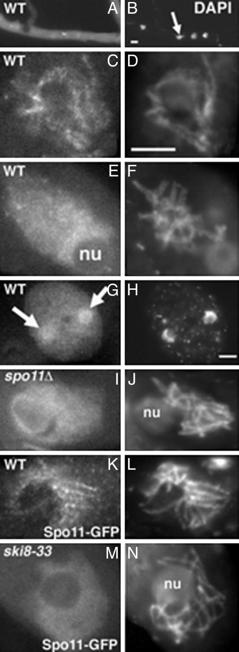
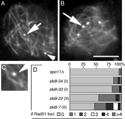
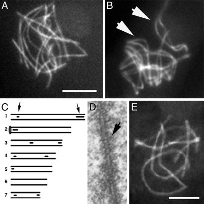
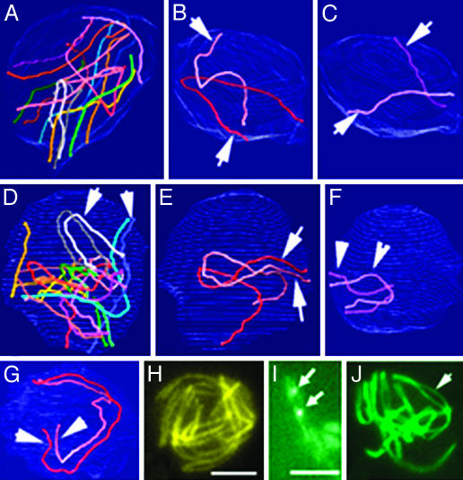
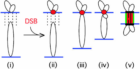
Ski8p is implicated in degradation of non-poly(A) and double-stranded RNA, and in meiotic DNA recombination. We have identified the Sordaria macrospora SKI8 gene. Ski8p is cytoplasmically localized in all vegetative and sexual cycle cells, and is nuclear localized, specifically in early-mid-meiotic prophase, in temporal correlation with Spo11p, the meiotic double-strand break (DSB) transesterase. Localizations of Ski8p and Spo11p are mutually interdependent. ski8 mutants exhibit defects in vegetative growth, entry into the sexual program, and sporulation. Diverse meiotic defects, also seen in spo11 mutants, are diagnostic of DSB absence, and they are restored by exogenous DSBs. These results suggest that Ski8p promotes meiotic DSB formation by acting directly within meiotic prophase chromosomes. Mutant phenotypes also divide meiotic homolog juxtaposition into three successive, mechanistically distinct steps; recognition, presynaptic alignment, and synapsis, which are distinguished by their differential dependence on DSBs.


Figures

Fig. 1.
Fig. 1.
Ski8 localization. (AH) WT. (A) Mycelium with bright Ski8-GFP signal. (B) Corresponding DAPI with four nuclei (arrow). (C) Late leptotene nucleus with chromatin-associated Ski8-GFP. (D) Corresponding DAPI. (E) Late pachytene nucleus with diffuse Ski8 signal. (F) Corresponding DAPI. (G) Ascospore with two nuclei (arrows) stained by Ski8-GFP. Note also the bright cytoplasmic staining when compared with the ascus staining in E. (H) Corresponding DAPI. (I) Ski8-GFP staining in a spo11Δ prophase nucleus. (J) Corresponding DAPI. (K) Spo11-GFP staining in a WT bouquet nucleus. (L) Corresponding DAPI. (M) Spo11-GFP staining in a ski8-33 leptotene nucleus. (N) Corresponding DAPI. nu, nucleolus. (Bar, 5 μm.)
Fig. 2.
Fig. 2.
Rad51p foci in ski8 leptotene nuclei double-stained by Spo76-GFP and anti-Rad51 antibodies. (A) ski8-33 with two foci (arrows). (B) ski8-7 with six foci (arrow points to brighter dots). (C) Enlargement of upper Rad51 focus in A.(D) Distribution of focus numbers per nucleus in ski8 mutants and spo11Δ. (Bar, 5 μm.)
Fig. 3.
Fig. 3.
Pairing and synapsis phenotypes of ski8 mutants. (A) Type I ski8-33 nucleus with only unpaired homologs. (B) Type II ski8-22 nucleus with aligned homologs (arrows point to two bivalents). (C) EM-reconstructed ski8-7 nucleus. Six of the seven homolog pairs show SC initiation sites (arrows point to bivalent 1, gray circle; bivalent 2 ends in nucleolus). (D)AEof ski8-22 is doubled and striated (arrow). (E) ski8-7 nucleus with seven synapsed bivalents. (Bars, 5μm for LM; 1 μm for EM.)
Fig. 4.
Fig. 4.
Progressive colocalization of homologs in WT. (A–G) 3D EM reconstructions of three serially sectioned nuclei. (A–C) Early leptotene nucleus. (A) The seven homologs are represented with matching colors. (B) Homolog pair 1 (arrows). (C) Pair 7 (arrows). These two pairs are easy to distinguish by their size. They show no sign of pairing. (D–F) Mid-leptotene nucleus with partially aligned homologs (arrows). (D) Overview. (E) Pair 1 shows only telomere alignment (arrows). (F) The small pair 7 is completely aligned. (G) At late leptotene, all homologs are completely aligned, illustrated here by pair 1 (arrows). (H) LM of equivalent nucleus (axes are stained by Spo76-GFP). (I) Two pairs of homologs (stained by Spo76-GFP) show Rad51-GFP foci (arrows) between the aligned axes. (J) Zygotene nucleus with partially synapsed bivalents (arrow) and telomeres grouped into a bouquet configuration. (Bars, 5 μm for LM and 1 μm for EM.)
Fig. 5.
Fig. 5.
Stages of homolog juxtaposition in mutants. (A and B) EM reconstruction of a ski8-33 (type I) nucleus. Homologs of pair 1 (A) and 7 (B) show only long distance partial recognition segments (arrows). (C–K) Spo76-GFP staining. (C) ski8-7 nucleus with clear homologous alignment (arrows point to three pairs). Type I ski8-34 (D) shows no alignment, whereas type II ski8-22 (E) does (arrows point to two pairs). (F and G) Spo76-GFP and Rad51-GFP (arrows) double-stained ski8-7 (F) and ski8-22 (G) nuclei. (H and I) Exposure at 200 Gy. (H) Complete synapsis in ski8-7 after 8 h. (I) Homolog alignment in ski8-33 (arrows) after 6 h. (J and K) Exposure at 100 Gy. (J) ski8-33 nucleus with aligned homologs (arrows). (K) ski8-22 nucleus shows a mixture of synapsed (arrowhead), aligned (thin arrow), and still roughly aligned (large arrow) homologs. (Bars, 5 μm for LM and 1 μm for EM.)
Fig. 6.
Fig. 6.
Model for DSB-mediated presynaptic axis coalignment. Homologs linked by DSB-independent homolog recognition (i) undergo a DSB. A DSB within an axis-associated recombination complex (red ball) captures the homologous sequence of a partner chromatin loop (ii) and reels the partner axis into close range by tracking along the partner loop (iii and iv), thus permitting formation of an interaxis structural bridge overlaid with a nascent DSB/ partner/RecA homolog complex (v).

References

    1. Zickler, D. & Kleckner, N. (1999) Annu. Rev. Genet. 33 603–754. - PubMed
    1. Lee, J. Y. & Orr-Weaver, T. L. (2001) Annu. Rev. Cell Dev. Biol. 17 753–777. - PubMed
    1. Keeney, S. (2001) Curr. Top. Dev. Biol. 52 1–53. - PubMed
    1. Paques, F. & Haber, J. E. (1999) Microbiol. Mol. Biol. Rev. 63 349–404. - PMC - PubMed
    1. Ridley, S. P., Sommer, S. S. & Wickner, R. B. (1984) Mol. Cell. Biol. 4 761–770. - PMC - PubMed

Publication types

LinkOut - more resources