. 2004 Jan;15(1):189-96.
doi: 10.1091/mbc.e03-08-0605. Epub 2003 Oct 17.

Disease-related myotubularins function in endocytic traffic in Caenorhabditis elegans

Affiliations

Disease-related myotubularins function in endocytic traffic in Caenorhabditis elegans

Hope Dang et al. Mol Biol Cell. 2004 Jan.

Abstract

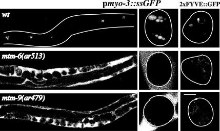
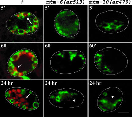
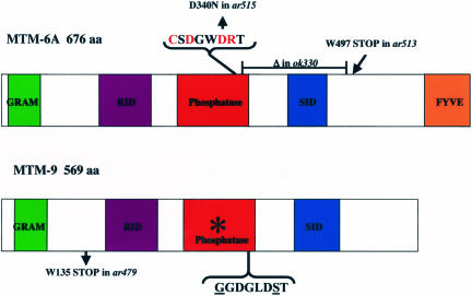
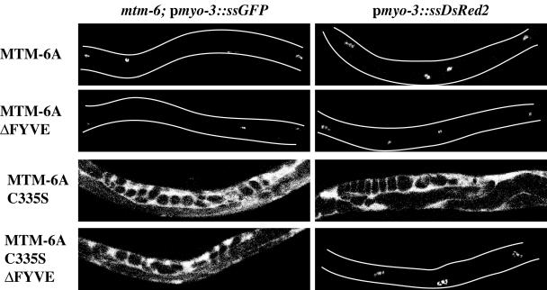
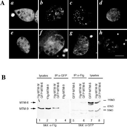
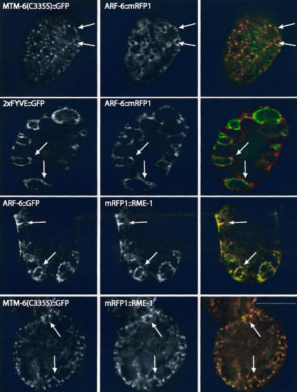
MTM1, MTMR2, and SBF2 belong to a family of proteins called the myotubularins. X-linked myotubular myopathy, a severe congenital disorder characterized by hypotonia and generalized muscle weakness in newborn males, is caused by mutations in MTM1 (Laporte et al., 1996). Charcot-Marie-Tooth types 4B1 and 4B2 are severe demyelinating neuropathies caused by mutations in MTMR2 (Bolino et al., 2000) and SBF2/MTMR13 (Senderek et al., 2003), respectively. Although several myotubularins are known to regulate phosphoinositide-phosphate levels in cells, little is known about the actual cellular process that is defective in patients with these diseases. Mutations in worm MTM-6 and MTM-9, myotubularins belonging to two subgroups, disorganize phosphoinositide 3-phosphate localization and block endocytosis in the coelomocytes of Caenorhabditis elegans. We demonstrate that MTM-6 and MTM-9 function as part of a complex to regulate an endocytic pathway that involves the Arf6 GTPase, and we define protein domains required for MTM-6 activity.


Figures

Figure 1.
Figure 1.
Endocytosis defects of mtm mutant strains. Shown are confocal images of wild-type, mtm-6(ar513), and mtm-9(ar479) worms either expressing pmyo-3::ssGFP (left two columns) or 2 × FYVE::GFP in their coelomocytes (right column). Worms carrying pmyo-3::ssGFP secrete GFP from their muscles into the body cavity. The coelomocytes (bright spots in the wild-type worms) endocytose the GFP from the body cavity. Defects in endocytosis result in GFP accumulating in the body cavity. The left column is a low-magnification image; the middle and last column are higher magnification images showing individual coelomocytes. Outlines of worms and coelomocytes are drawn when needed. Bar, 5 μm for the high-magnification images.
Figure 2.
Figure 2.
Time course of uptake of BSA-Rhod by coelomocytes expressing RME-8::GFP in wild-type, mtm-6(ar513), or mtm-9(ar479) worms. The images are overlays of GFP (green) and rhodamine (red) fluorescence images taken with a confocal microscope. The time the pictures were taken after injection are indicated. The large arrow indicates concentrations of the BSA-Rhod in endosomes labeled with RME-8::GFP. Small arrows indicate lysosomes that are not labeled with RME-8::GFP. The arrowheads indicate extremely faint rhodamine staining in the coelomocytes of the mutant worms after 24 h of uptake. Outlines of worms and are drawn. Bar, 5 μm in all images.
Figure 3.
Figure 3.
MTM-6– and MTM-9–predicted proteins and mutant alleles. Schematic representation of the proteins, and their various domains, predicted from the cDNA sequences. The catalytic amino acids of the phosphatase domain of MTM-6 are indicated in red and the corresponding substituted amino acids of MTM-9 are underlined. The asterisk denotes that the phosphatase domain of MTM-9 lacks residues thought to be required for activity. The predicted changes in the various mutant alleles are shown. mtm-6(ok330) and mtm-9(ar479) represent the molecular null alleles. All the alleles of mtm-6 gave identical phenotypes.
Figure 4.
Figure 4.
Effect of MTM-6 alleles on endocytosis by coelomocytes. Shown are low-magnification confocal images of mtm-6(ar513); pmyo-3::ssGFP or of pmyo-3::ssDsRed2 worms carrying transgenes expressing GFP fusions of various forms of MTM-6A. Bright spots in the worms are coelomocytes normally endocytosing the fluid-phase marker. Bright fluorescence in the body cavity indicates absence of endocytosis. Outlines of worms are drawn when needed.
Figure 5.
Figure 5.
Subcellular localization and interaction of MTM-6A and MTM-9. (A) High-magnification confocal images of the GFP fusion protein expressed in the coelomocytes of the following strains (Ex signifies the transgenic array from which the indicated protein is expressed): a, +; Ex[MTM-6A::GFP]; b, +; Ex[MTM-6A(C335S):: GFP]; c, mtm-9(ar479); Ex[MTM-6A(C335S)::GFP]; d, rme-1(b1045); Ex[MTM-6A(C335S)::GFP]; e, +; Ex[MTM-6AΔFYVE::GFP]; f, +; Ex[MTM-6A (C335S)ΔFYVE::GFP]; g, +; Ex[MTM9::GFP]; and h, Ex[MTM-6A(C335S)]; Ex[MTM9::GFP]. Bar, 5 μm in all images. (B) Coimmunoprecipitation of GFP-MTM-6A and Flg-MTM-9 expressed in human 293 cells. Antibodies against GFP or Flg were used to immunoprecipitate the corresponding fusion protein indicated above the gel. Blots were probed for the presence of the other fusion protein by using the antibodies indicated below the gel.
Figure 6.
Figure 6.
Deconvolution images of wild-type worms expressing the indicated proteins from transgenes. Panels in the right column are overlays of the corresponding GFP (green) and mRFP1 (red) images. Arrows indicate some of the membranes to which both proteins colocalize. Bar, 5 μm in all images.

References

    1. Berger, P., Bonneick, S., Willi, S., Wymann, M., and Suter, U. (2002). Loss of phosphatase activity in myotubularin-related protein 2 is associated with Charcot-Marie-Tooth disease type 4B1. Hum. Mol. Genet. 11, 1569–1579. - PubMed
    1. Blondeau, F., Laporte, J., Bodin, S., Superti-Furga, G., Payrastre, B., and Mandel, J.L. (2000). Myotubularin, a phosphatase deficient in myotubular myopathy, acts on phosphatidylinositol 3-kinase and phosphatidylinositol 3-phosphate pathway. Hum. Mol. Genet. 9, 2223–2229. - PubMed
    1. Bolino, A., et al. (2000). Charcot-Marie-Tooth type 4B is caused by mutations in the gene encoding myotubularin-related protein-2. Nat. Genet. 25, 17–19. - PubMed
    1. Boshans, R.L., Szanto, S., van Aelst, L., and D'Souza-Schorey, C. (2000). ADP-ribosylation factor 6 regulates actin cytoskeleton remodeling in coordination with Rac1 and RhoA. Mol. Cell. Biol. 20, 3685–3694. - PMC - PubMed
    1. Brenner, S. (1974). The genetics of Caenorhabditis elegans. Genetics 77, 71–94. - PMC - PubMed

Publication types

MeSH terms

LinkOut - more resources