. 2003 Dec;14(12):4931-46.
doi: 10.1091/mbc.e03-06-0419. Epub 2003 Oct 17.

Interactions between centromere complexes in Saccharomyces cerevisiae

Affiliations

Interactions between centromere complexes in Saccharomyces cerevisiae

Vladimir S Nekrasov et al. Mol Biol Cell. 2003 Dec.

Abstract

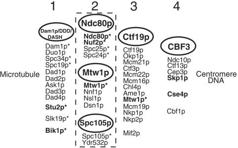
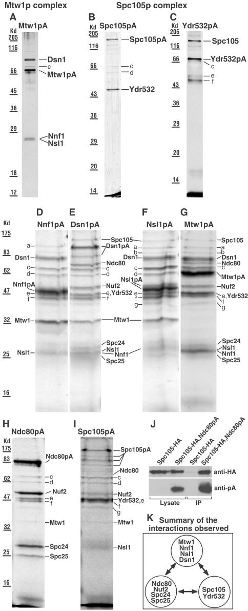
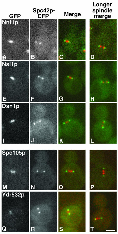
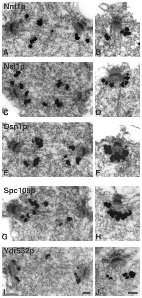
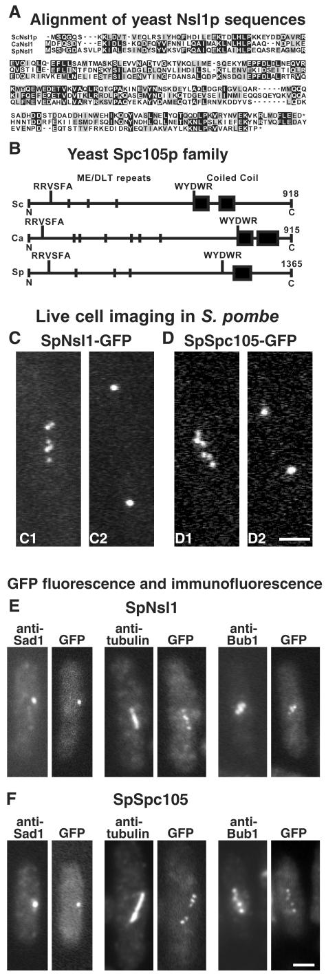
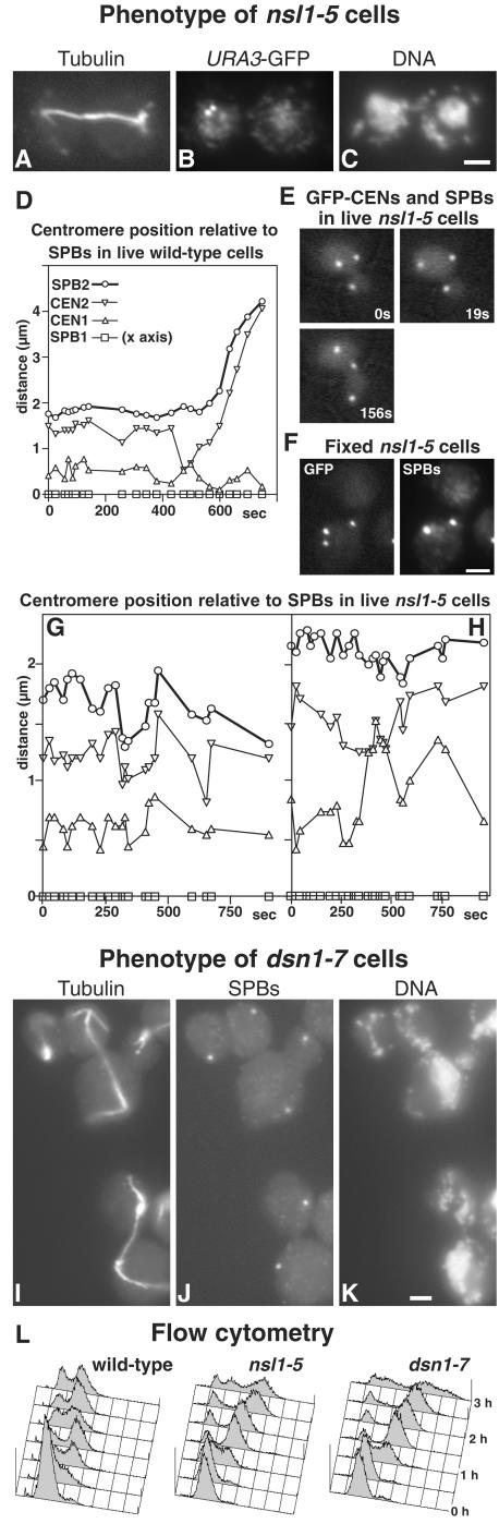
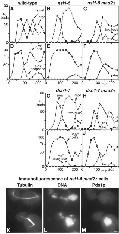
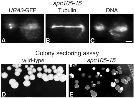
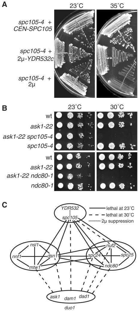
We have purified two new complexes from Saccharomyces cerevisiae, one containing the centromere component Mtw1p together with Nnf1p, Nsl1p, and Dsn1p, which we call the Mtw1p complex, and the other containing Spc105p and Ydr532p, which we call the Spc105p complex. Further purifications using Dsn1p tagged with protein A show, in addition to the other components of the Mtw1p complex, the two components of the Spc105p complex and the four components of the previously described Ndc80p complex, suggesting that all three complexes are closely associated. Fluorescence microscopy and immunoelectron microscopy show that Nnf1p, Nsl1p, Dsn1p, Spc105p, and Ydr532p all localize to the nuclear side of the spindle pole body and along short spindles. Chromatin immunoprecipitation assays show that all five proteins are associated with centromere DNA. Homologues of Nsl1p and Spc105p in Schizosaccharomyces pombe also localize to the centromere. Temperature-sensitive mutations of Nsl1p, Dsn1p, and Spc105p all cause defects in chromosome segregation. Synthetic-lethal interactions are found between temperature-sensitive mutations in proteins from all three complexes, in agreement with their close physical association. These results show an increasingly complex structure for the S. cerevisiae centromere and a probable conservation of structure between parts of the centromeres of S. cerevisiae and S. pombe.


Figures

Figure 10.
Figure 10.
Possible groupings of S. cerevisiae centromere and kinetochore components. Components in bold type have known human homologues. Groups 1 and 2 contain components (with asterisks) that have been detected in spindle pole preparations by MALDI mass spectrometry (Wigge et al., 1998; Wigge and Kilmartin, 2001). Group 1 components associate laterally with microtubules and have also been detected at the centromere by ChIP assay. Group 2 components localize to the centromere only; the dashed line symbolizes the close association of these complexes. Nearly all group 3 components (Meluh and Koshland, 1995; Cheeseman et al., 2002a; Pot et al., 2003) have been localized to the centromere only and have not been detected in spindle pole preparations (except for Mtw1p; see text). Group 4 components bind centromere DNA directly as judged by band shift assay. References for most of these components can be found in the Introduction.
Figure 1.
Figure 1.
(A–I) SDS gels of the complexes isolated from strains (names in brackets) containing the indicated protein A (pA)-tagged proteins. (A and G) Mtw1p (MSY5), (B and I) Spc105p (PWY282), (C) Ydr532p (MSY48), (D) Nnf1p (VNY34), (E) Dsn1p (VNY38), (F) Nsl1p (VNY32), and (H) Ndc80p (PWY350). Gels A–C were from the first fraction off the columns and gels D–I from the second fraction, which usually contains more material but also more contaminants. All the gels were Coomassie-stained except A and C, which were silver-stained. In gels D–I, the sample wells were loaded as fully as possible, which caused some distortion in the band shape. In E, the lower band labeled Dsn1pA was a proteolytic fragment since it contained peptides from both Dsn1p and protein A. In F, no further proteins were identified in the Nnf1p region of this gel. Bands a-g were identified as Rpn1p (a), Sen3p (b, only identified in gels F and G), Ssa1p, Ssa2p, or both (c), Hsp60p (d), Tef2p (e), Ydj1p (f), and Scj1p (g). Rpn1p and Sen3p are proteosome components, Ssa1p, Ssa2p, Hsp60p, Ydj1p, and Scj1p are heat shock factors, and Tef2p is elongation factor. Further information on these proteins can be found at http://www.yeastgenome.org/. It seems likely that all of these are contaminants since they have been found in isolations with noncentromeric proteins coupled to protein A (our unpublished results), and most are absent from the cleaner gels (A–C). (J) Immunoblot of immunoprecipitates (IP) obtained as described in MATERIALS AND METHODS using strains containing HA-tagged Spc105p only (VNY290), or both HA-tagged Spc105p and protein A-tagged Ndc80p (VNY296). This shows that upon enriching for Ndc80pA, trace amounts of Spc105-HA can be detected. (K) Summary of the interactions observed.
Figure 2.
Figure 2.
Localization of GFP-tagged proteins in relation to the SPBs (imaged with Spc42p-CFP) in unfixed cells of the indicated strains. The left three columns show the GFP (red), CFP (green) and merged images for a cell of each strain with a short spindle. The right column shows the merged image for a cell with a longer spindle; note the splitting of the GFP image. (A–D) Nnf1p (JK1694), (E–H) Nsl1p (JK1682), (I–L) Dsn1p (JK1683), (M–P) Spc105p (JK1679), and (Q–T) Ydr532p (JK1676). There is a small amount of carryover of the GFP signal into the CFP channel. Bar, 2 μm.
Figure 3.
Figure 3.
Immuno-EM of GFP-tagged proteins in the indicated strains. (A and B) Nnf1p (VNY3), (C and D) Nsl1p (VNY50), (E and F) Dsn1p (VNY28), (G and H) Spc105p (MSY52), and (I and J) Ydr532p (JK1495). Staining is seen between SPBs in short spindles (left side) and in association with the nuclear face in all other SPBs (right side). Bar, 0.1 μm.
Figure 4.
Figure 4.
ChIP assays on protein A-tagged ndc10-1 strains and on the wild-type strain K699 as a negative control. Assays were carried out at 23°C (left), where the centromere is intact in ndc10-1 cells, and at 37°C (right), where the centromere is dissociated. Multiplex PCR was used to detect CEN3 and two regions 4 kb on either side. P is the pellet after enrichment with IgG Sepharose, and T, 1% of the total input, is the starting material. From left to right, the strains used were JK1670, JK1673, JK1667, JK1669, JK1664, and K699.
Figure 5.
Figure 5.
(A) Alignment of Nsl1p sequences from S. cerevisiae (Sc), C. albicans (Ca), and S. pombe (Sp); the last nine amino acids of the Candida homologue have been omitted. Black and gray boxes indicate identical and similar residues, respectively. (B) Schematic view of the Spc105p homologues from the same three species. ME/DLT repeats are shown by short vertical lines, and coiled-coil domains found by Paircoil (Berger et al., 1995) are shown by black boxes. (C and D) Localization of GFP-tagged SpNsl1 (C1 and C2) and SpSpc105 (D1 and D2) in live S. pombe cells. Long cells in which the GFP spots had begun to split, indicating entry into mitosis, were followed, and images were recorded when these cells were about to complete anaphase A (C1 and D1). These images show between five and six dots, which then coalesced into two separated dots several minutes later as the cells entered anaphase B (C2 and D2). (E and F) Fluorescence of GFP compared with immunofluorescence with anti-Sad1, antitubulin, and anti-HA-tagged SpBub1 is shown in E for SpNsl1 (strain JK1636) and in F for SpSpc105 (strain JK1627). Bars, 2 μm.
Figure 6.
Figure 6.
Phenotypes of nsl1-5 (A–C and E–H) and dsn1-7 (I–K) mutants, and of a wild-type control (D); flow cytometry for the three strains is also shown (L). Cells were synchronized in G1 with α-factor at 23°C and released at 36°C. (A–C) An nsl1-5 cell in which the URA3 locus is labeled with GFP (strain VNY287) was fixed during anaphase (3 h after release) and processed for immunofluorescence with antitubulin (A), and anti-GFP (B), and stained with DAPI (C). Chromosome V has failed to segregate. (D–H) Imaging of GFP-labeled SPBs and CEN5 in wild-type (D, strain K8572) and nsl1-5 cells (E–H, strain VNY122). (D) A wild-type cell underwent centromere pairing and passed through anaphase during the period of observation. (E) An nsl1-5 cell where the centromeres failed to split. (F) A fixed nsl1-5 cell was processed for immunofluorescence using anti-GFP and anti-Tub4p to identify SPBs. Centromeres (the lowest GFP spot) have not split. (G) One of the majority of nsl1-5 cells, in which the centromeres split but failed to pair during the period of observation. (H) An nsl1-5 cell in which pairing occurred. Symbols in G and H are the same as in D. (I–K) dsn1-7 cells (strain VNY96) were processed for immunofluorescence 2.5 h after release using antitubulin (I), anti-Tub4p for SPBs (J), and DAPI (K). Two anaphase cells that have failed to segregate DNA and one aploid cell (top left) are shown. (L) Flow cytometry of wild-type (K699), nsl1-5 (VNY72), and dsn1-7 (VNY96) cells. Bars, 2 μm.
Figure 7.
Figure 7.
The arrest of nsl1-5 and dsn1-7 mutants during mitosis is Mad2p-dependent. (A–J) Wild-type (A and D; K6445), nsl1-5 (B and E; VNY130), nsl1-5 mad2Δ (C and F; VNY318), dsn1-7 (G and I; VNY116), and dsn1-7 mad2Δ (H and J; VNY283) cells were synchronized in G1 with α-factor and released at 36°C. The percentage of cells with small buds, large buds, or two buds was plotted against time (A–C, G, and H), as were the percentages of Pds1p-positive cells and Pds1p-negative cells containing anaphase spindles as determined by immunofluorescence (D–F, I and J). (K–M) Immunofluorescence of nsl1-5 mad2Δ cells stained with antitubulin (K), DAPI (L), and antimyc for Pds1p (M). Pds1p-negative cells with anaphase spindles show unequal DNA segregation. Bar, 2 μm.
Figure 8.
Figure 8.
spc105-15 cells are defective in chromosome segregation. (A–C) spc105-15 cells in which the URA3 locus is labeled with GFP (strain VNY203) were synchronized in G1 with α-factor, released at 36°C for 2 h, fixed, and stained with anti-GFP (A), antitubulin (B), and DAPI for DNA (C). (D and E) Colony-sectoring assay for chromosome loss in wild-type (D; strain K7049) and spc105-15 (E; strain VNY198) cells carrying the CFIII mini-chromosome after incubation on low adenine YEPD for 3 d at 30°C. The red colonies (dark in the figure) and sectors show chromosome loss. Bar, 2 μm.
Figure 9.
Figure 9.
Genetic interactions within and between the Spc105p, Mtw1p, Ndc80p, and Dam1p/DDD/DASH complexes. (A) Suppression of spc105-4 by overexpression of Ydr532p. Strains were MSY150, MSY151, and MSY152. (B) Examples of the synthetic growth defects at 30°C between mutations in the different complexes. (C) A summary of the interactions found. Solid lines show synthetic-lethal interactions at 23°C, dashed lines show synthetic growth defects with lethality at 30°C (see panel B), the dotted line shows dosage suppression (see panel A), and the absence of a line indicates that no genetic interaction was found. All possible combinations between the different mutations were tested; however, the YDR532c dosage suppression was only tested in the spc105 alleles. The synthetic-lethal interactions within the Ndc80p and Mtw1p complexes have been described previously (Wigge and Kilmartin, 2001; Euskirchen, 2002). The ask1, dam1, and dad1 alleles are not distinguished because they gave similar results in all the crosses tested, with the exception of the dam1-31 mtw1-11 cross, in which no interaction was found. duo1 is excluded from the ask1, dad1, and dam1 group because duo1-61 showed no genetic interactions with any of the other alleles. Strains used were JK1121, JK1504, MSY72, MSY79, MSY87, MSY90, PWY483, PWY754, VNY69, VNY87, VNY96, and VNY162.

References

    1. Adams, A.E.M., and Pringle, J.R. (1984). Relationship of actin and tubulin distribution to bud growth in wild type and morphogenetic-mutant Saccharomyces cerevisiae. J. Cell Biol. 98, 934–945. - PMC - PubMed
    1. Adams, I.R., and Kilmartin, J.V. (1999). Localization of core spindle pole body (SPB) components during SPB duplication in Saccharomyces cerevisiae. J. Cell Biol. 145, 809–823. - PMC - PubMed
    1. Aebersold, R., and Mann, M. (2003). Mass spectrometry-based proteomics. Nature 422, 198–207. - PubMed
    1. Bähler, J., Wu, J.-Q., Longtine, M.S., Shah, N.G., McKenzie, A., III, Steever, A.B., Wach, A., Philippsen, P., and Pringle, J.R. (1998). Heterologous modules for efficient and versatile PCR-based gene targeting in Schizosaccharomyces pombe. Yeast 14, 943–951. - PubMed
    1. Berger, B., Wilson, D.B., Wolf, E., Tonchev, T., Milla, M., and Kim, P.S. (1995). Predicting coiled coils by use of pairwise residue correlations. Proc. Natl. Acad. Sci. USA 92, 8259–8263. - PMC - PubMed

Publication types

MeSH terms

Substances