. 2003 Dec;14(12):4920-30.
doi: 10.1091/mbc.e03-04-0264. Epub 2003 Oct 17.

The adhesin Hwp1 and the first daughter cell localize to the a/a portion of the conjugation bridge during Candida albicans mating

Affiliations

The adhesin Hwp1 and the first daughter cell localize to the a/a portion of the conjugation bridge during Candida albicans mating

Karla J Daniels et al. Mol Biol Cell. 2003 Dec.

Abstract

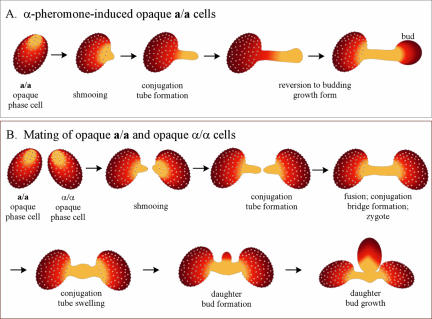
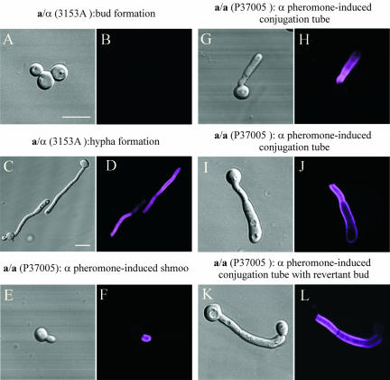
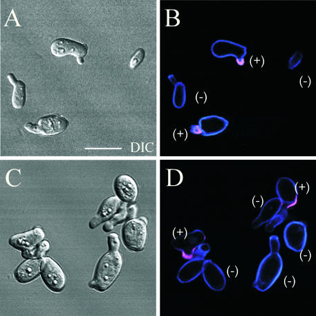
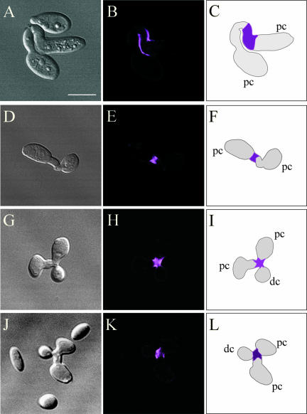
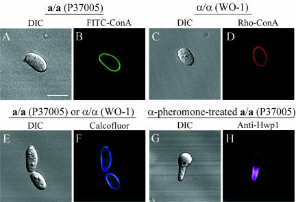
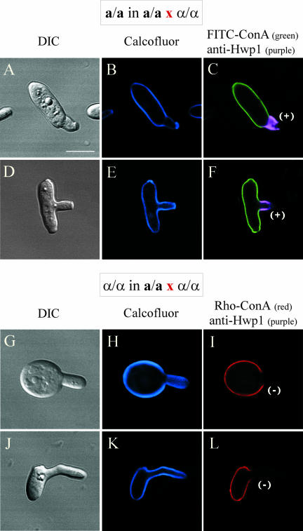
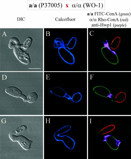
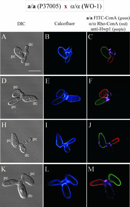
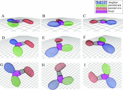
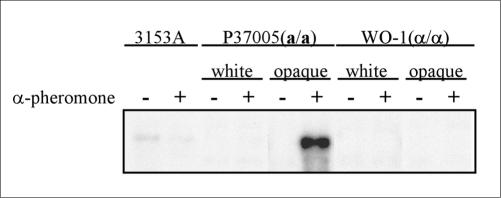
The cell wall protein Hwp1 was originally demonstrated to be expressed exclusively in hyphae of Candida albicans and cross-linked to human epithelium by mammalian transglutaminase. Hwp1 is expressed on the walls of hyphae formed by a/alpha, a/a, and alpha/alpha cells. Hence, it is expressed on hyphae independently of mating type. However, Hwp1 is selectively expressed on the wall of conjugation tubes formed by a/a cells, but not alpha/alpha cells, in the mating process. This was demonstrated in all possible crosses between four unrelated natural a/a strains and four unrelated alpha/alpha strains. In zygotes, Hwp1 is restricted to that portion of the wall of the conjugation bridge contributed by the a/a parent cell. Hwp1 staining further revealed that the first daughter bud that emerges from the conjugation bridge does so from the a/a-contributed portion. Hwp1 expression and localization during the mating process is, therefore, mating type specific, opaque phase specific, and alpha-pheromone induced. These results indicate that the mating type-specific contributions to the conjugation bridge during the mating process in C. albicans are qualitatively and functionally distinct and that the a/a portion of the bridge, which selectively contains Hwp1, bears the first daughter cell in the mating process.


Figures

Figure 1.
Figure 1.
Models of α-pheromone-induced conjugation tube formation and reversion to the budding growth form (A), and a/a × α/α mating in C. albicans (B).
Figure 2.
Figure 2.
Anti-Hwp1 antibody staining reveals that Hwp1 is expressed in α-pheromone-induced a/a conjugation tubes as well as serum-induced hyphae. Cells are stained with anti-Hwp1 antibody. (A and B) White budding cell of the a/α strain 3153A. (C and D) Hypha formation of the a/α strain 3153A. (E and F) α-Pheromone-induced shmoo formation of the a/a strain P37005. (G and H and I and J) α-Pheromone-induced conjugation tube formation. (K and L) Reversion of a conjugation tube to the budding growth form at the tube apex. Each pair of images includes a DIC image and a confocal optical section through the center of the cell. Hwp1 stains purple. Bar, 5 μm.
Figure 3.
Figure 3.
Anti-Hwp1 antibody stains only half of the shmoos and conjugation tubes of a/a × α/α mating cultures. Cells were stained with Calcofluor, which stains all cell wall blue, and anti-Hwp1 antibody, which stains Hwp1 purple. Each pair of images includes a DIC image and a confocal image taken through the center of cells. Bar, 5 μm.
Figure 4.
Figure 4.
Anti-Hwp1 antibody stains only half of each conjugation bridge, which represents the portion of the bridge from which the first daughter bud emerges. Each of the four sets of images includes a DIC image, a confocal optical section, and a diagram of the staining pattern. Bar, 5 μm.
Figure 5.
Figure 5.
Multiple staining procedure. (A and B) DIC and FITC-ConA staining (green) of an a/a cell. (C and D) DIC and rhodamine-ConA staining (red) of an α/α cell. (E and F) DIC and calcofluor-staining of a/a or α/α cells. (G and H) DIC and anti-Hwp1 antibody-staining of an early conjugation tube formed by an α-pheromone-induced a/a cell. In the procedure, a/a and α/α parent cells are vitally stained with FITC-ConA and rhodamine-ConA, respectively, and then allowed to mate. The embryos are then fixed and stained with anti-Hwp1 antibody plus secondary antibody and calcofluor. Bar, 5 μm.
Figure 6.
Figure 6.
The multiple staining procedure demonstrates that in an a/a × α/α mating culture, only conjugation tubes formed by a/a cells contain Hwp1. FITC-ConA–stained (green) a/a cells and rhodamine-ConA–stained (red) α/α cells were mixed for 2 or 6 h and then fixed and stained with anti-Hwp1 antibody and calcofluor. (A–C; D–F) a/a cells with tubes. (G–I; J–L) α/α cells with tubes. +, staining of HWP1; –, no staining of Hwp1. Bar, 5 μm.
Figure 7.
Figure 7.
The multiple staining procedure demonstrates that only the a/a contribution to the conjugation bridge contains Hwp1. FITC-ConA–stained (green) a/a cells and rhodamine-ConA–stained (red) α/α cells were mixed for 18 h and then fixed and stained with anti-Hwp1 antibody and calcofluor. (A–C; D–F; G–I) Individual fusants before bud formation.. Bar, 5 μm.
Figure 8.
Figure 8.
The multiple staining procedure demonstrates that daughter cells form only on the a/a contribution to the bridge. FITC-ConA–stained (green) a/a cells and rhodamine-ConA–stained (red) α/α cells were mixed for 18 h and then fixed and stained with anti-Hwp1 antibody and calcofluor. (A–C; D–F; G–I; J–L) Individual zygotes. Bar, 5 μm.
Figure 9.
Figure 9.
3D-DIAS faceted reconstruction of a zygote demonstrates that the first bud is formed from the a/a contribution of the bridge. The a/a parent cell is color-coded green, the α/α parent cell is color-coded red, the Hwp1-containing portion of the bridge is color-coded purple, the portion of the bridge lacking Hwp1 is color-coded black, and the daughter cell is color-coded blue. (A–C) Zygote viewed at a 15° angle, rotated in the x,y-axis. (D–F) Zygote viewed at 45°, rotated in the x,y-axis. (G–I) Zygote viewed at 90°, rotated in the x,y-axis.
Figure 10.
Figure 10.
A Northern blot analysis of HWP1 expression demonstrates that HWP1 transcription is induced only in opaque a/a cells by α-pheromone.

References

    1. Anderson, J.M., and Soll, D.R. (1987). Unique phenotype of opaque cells in the white-opaque transition of Candida albicans. J. Bacteriol. 169, 5579–5588. - PMC - PubMed
    1. Bedell, G., and Soll, D.R. (1979). The effects of low concentrations of zinc on the growth and dimorphism of Candida albicans: evidence for zinc resistant and zinc sensitive pathways for mycelium formation. Infect. Immun. 26, 348–354. - PMC - PubMed
    1. Buffo, J., Herman, M., and Soll, D.R. (1984). A characterization of pH-regulated dimorphism in Candida albicans. Mycopathologia 85, 21–30. - PubMed
    1. Casamayor, A., and Snyder, M. (2002). Bud site selection and cell polarity in budding yeast. Curr. Opin. Microbiol. 5, 179–186. - PubMed
    1. Cole, G.M., and Reed, S.I. (1991). Pheromone-induced phosphorylation of a G protein beta subunit in S. cerevisiae is associated with an adaptive response to mating pheromone. Cell 64, 703–716. - PubMed

Publication types

MeSH terms

LinkOut - more resources