. 2003 Dec;14(12):5082-8.
doi: 10.1091/mbc.e03-04-0246. Epub 2003 Oct 17.

Dissection of the Ascaris sperm motility machinery identifies key proteins involved in major sperm protein-based amoeboid locomotion

Affiliations

Dissection of the Ascaris sperm motility machinery identifies key proteins involved in major sperm protein-based amoeboid locomotion

Shawnna M Buttery et al. Mol Biol Cell. 2003 Dec.

Abstract

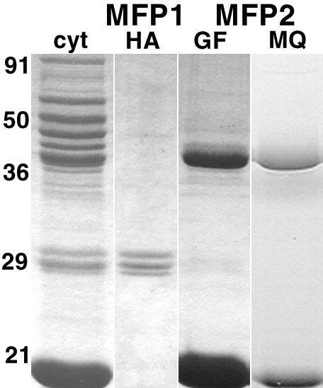
Although Ascaris sperm motility closely resembles that seen in many other types of crawling cells, the lamellipodial dynamics that drive movement result from modulation of a cytoskeleton based on the major sperm protein (MSP) rather than actin. The dynamics of the Ascaris sperm cytoskeleton can be studied in a cell-free in vitro system based on the movement of plasma membrane vesicles by fibers constructed from bundles of MSP filaments. In addition to ATP, MSP, and a plasma membrane protein, reconstitution of MSP motility in this cell-free extract requires cytosolic proteins that orchestrate the site-specific assembly and bundling of MSP filaments that generates locomotion. Here, we identify a fraction of cytosol that is comprised of a small number of proteins but contains all of the soluble components required to assemble fibers. We have purified two of these proteins, designated MSP fiber proteins (MFPs) 1 and 2 and demonstrated by immunolabeling that both are located in the MSP cytoskeleton in cells and in fibers. These proteins had reciprocal effects on fiber assembly in vitro: MFP1 decreased the rate of fiber growth, whereas MFP2 increased the growth rate.


Figures

Figure 1.
Figure 1.
Cytosol can be separated into less complex fractions that are competent to assemble fibers. (A) Coomassie-stained SDS-PAGE gel of cytosol, 25-40%, and 10-25% ammonium sulfate fractions and the SP-Sepharose-bound subfraction. Phase contrast micrographs of fibers assembled with vesicles, MSP and ATP in combination with cytosol (C), the 25-40% fraction (D), the 10-25% fraction (E), or the SP-Sepharose-bound subfraction (F). Bar, 5 μm. Two antibodies were used to screen the SP-Sepharose-bound fraction of cytosol in Western blots (B). Anti-Mr ∼38 kDa recognized a single band on Western blots. Monoclonal antibody 6F9 recognized a triplet of Mr ∼29 kDa.
Figure 2.
Figure 2.
Indirect immunofluorescence with antibodies against MFP1 and MFP2 labels the MSP cytoskeleton uniformly in vivo and in vitro. (A and D) Immunolabeling of a fixed, permeabilized sperm showing labeling throughout the fiber complexes that comprise the MSP cytoskeleton. (B and C and E and F) The paired immunofluorescence and differential interference contrast images of fibers assembled in vitro. In both cases, the fiber is labeled uniformly but its associated vesicle (arrows) is not. Bars, 10 μm.
Figure 3.
Figure 3.
Isolation of MFP1 and 2 for in vitro assays. Coomassie-stained gels of cytosol (cyt), the starting material for isolation of MFP1 and 2. MFP1, was separated by hydroxyapatite (HA) chromatography, whereas MFP2 was obtained by sequential gel permeation (GF) and anion exchange (MQ).
Figure 4.
Figure 4.
Effects of MFP1 and MFP2 on fiber growth rate at varying dilutions of S100 compared with that of dilution with KPM buffer alone. The mean rate of fiber growth with MFP1 added at 7 mg/ml (A) or MFP2 added at 14 mg/ml (B) to selected dilutions of S100. Each entry represents the mean of three trials, each including measurements from 10 to 15 fibers. The bars are the SEs of the means.
Figure 5.
Figure 5.
Effects of MFP2 are antagonistic to the effects of MFP1. The mean rate of fiber growth with addition of MFP1, MFP2, and anti-MFP1 singly and in pairwise combinations to 1:15 dilutions of S100. The rate-enhancing effect of MFP2 overcomes the rate-inhibiting effect of MFP1. Furthermore, combination of anti-MFP1 and MFP2 does not increase fiber growth rate more than either anti-MFP1 or MFP2 alone. Each point represents the average of two trials, each with 10-15 fibers. The error bars represent the SD from the means.
Figure 6.
Figure 6.
Perfusion assays confirm that MFP1 decreases and MFP2 increases fiber growth rate. (A) A control fiber grown in 1:10 diluted S100 and then perfused with ATP and cytosol (S100 with the vesicles removed by centrifugation) at the same dilution at the point indicated as “cytosol on.” The bars indicate the length of the fiber assembled 5 min before (to the right) and after (to the left) perfusion. The rate of growth was unchanged. (B) Fiber perfused with cytosol with MFP1 triplet at 7 mg/ml slowed substantially after perfusion. Note that the optical density of the segment grown in the presence of added MFP1 was lower than that in the segment grown before perfusion. (C) A similar fiber perfused with cytosol supplemented with MFP2 at 7 mg/ml. The added MFP2 increased the rate of fiber growth more than twofold without a change in optical density. Bar, 10 μm.
Figure 7.
Figure 7.
Predicted protein sequences of MFP1 and MFP2 and their C. elegans homologs. (A) Alignment of MFP1α (accession no. AY326285) and MFP1β (accession no. AY326286) with MSP domain protein 1 (MDP1; accession no. AY135488.1) from Ascaris and C. elegans C35D10.11 (Ce). (B) Alignment of MFP2 (accession no. AY326287) predicted protein sequence with C. elegans ZK265.3 (Ce). Proline-rich regions are shown in italics. (C) Alignment of the two domains (amino acids 3-101 and 176-278) of MFP2 demonstrates a 32% homology. In each alignment, underlined regions indicate peptides that were obtained from the native protein, asterisks represent identical residues, colons indicate highly conserved amino acids, and periods denote less conserved residues.

References

    1. Borisy, G.G., and Svitkina, T.M. (2000). Actin machinery: pushing the envelope. Curr. Opin. Cell Biol. 12, 104-112. - PubMed
    1. Holt, M.R., and Koffer, A. (2001). Cell motility: proline-rich proteins promote protrusions. Trends Cell Biol. 11, 38-46. - PubMed
    1. Italiano, J.E., Jr., Roberts, T.M., Stewart, M., and Fontana, C.A. (1996). Reconstitution in vitro of the motile apparatus from the amoeboid sperm of Ascaris shows that filament assembly and bundling move membranes. Cell 84, 105-114. - PubMed
    1. Italiano, J.E., Jr., Stewart, M., and Roberts, T.M. (2001). How the assembly dynamics of the nematode major sperm protein generate amoeboid cell motility. Int. Rev. Cytol. 202, 1-34. - PubMed
    1. Kay, B.K., Williamson, M.P., and Sudol, M. (2000). The importance of being proline: the interaction of proline-rich motifs in signaling proteins with their cognate domains. FASEB J. 14, 231-241. - PubMed

Publication types

Substances