

Analysis of the dynein-dynactin interaction in vitro and in vivo
- PMID: 14565986
- PMCID: PMC284810
- DOI: 10.1091/mbc.e03-01-0025
Analysis of the dynein-dynactin interaction in vitro and in vivo
Abstract
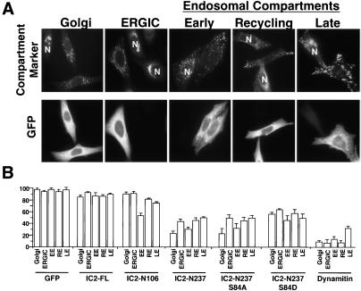
Cytoplasmic dynein and dynactin are megadalton-sized multisubunit molecules that function together as a cytoskeletal motor. In the present study, we explore the mechanism of dynein-dynactin binding in vitro and then extend our findings to an in vivo context. Solution binding assays were used to define binding domains in the dynein intermediate chain (IC) and dynactin p150Glued subunit. Transient overexpression of a series of fragments of the dynein IC was used to determine the importance of this subunit for dynein function in mammalian tissue culture cells. Our results suggest that a functional dynein-dynactin interaction is required for proper microtubule organization and for the transport and localization of centrosomal components and endomembrane compartments. The dynein IC fragments have different effects on endomembrane localization, suggesting that different endomembranes may bind dynein via distinct mechanisms.
Figures
Similar articles
-
Regulation of dynactin through the differential expression of p150Glued isoforms.J Biol Chem. 2008 Nov 28;283(48):33611-9. doi: 10.1074/jbc.M804840200. Epub 2008 Sep 22. J Biol Chem. 2008. PMID: 18812314 Free PMC article.
-
Recruitment of CG-NAP to the Golgi apparatus through interaction with dynein-dynactin complex.Genes Cells. 2007 Mar;12(3):421-34. doi: 10.1111/j.1365-2443.2007.01055.x. Genes Cells. 2007. PMID: 17352745
-
Preferentially localized dynein and perinuclear dynactin associate with nuclear pore complex proteins to mediate genomic union during mammalian fertilization.J Cell Sci. 2003 Dec 1;116(Pt 23):4727-38. doi: 10.1242/jcs.00784. J Cell Sci. 2003. PMID: 14600259
-
[Dynein and dynactin as organizers of the system of cell microtubules].Ontogenez. 2006 Sep-Oct;37(5):323-39. Ontogenez. 2006. PMID: 17066975 Review. Russian.
-
The role of the dynactin complex in intracellular motility.Int Rev Cytol. 1998;182:69-109. doi: 10.1016/s0074-7696(08)62168-3. Int Rev Cytol. 1998. PMID: 9522459 Review.
Cited by
-
Self-organization of stabilized microtubules by both spindle and midzone mechanisms in Xenopus egg cytosol.Mol Biol Cell. 2013 May;24(10):1559-73. doi: 10.1091/mbc.E12-12-0850. Epub 2013 Mar 20. Mol Biol Cell. 2013. PMID: 23515222 Free PMC article.
-
MINFLUX reveals dynein stepping in live neurons.Proc Natl Acad Sci U S A. 2024 Sep 17;121(38):e2412241121. doi: 10.1073/pnas.2412241121. Epub 2024 Sep 10. Proc Natl Acad Sci U S A. 2024. PMID: 39254993 Free PMC article.
-
Golgin160 recruits the dynein motor to position the Golgi apparatus.Dev Cell. 2012 Jul 17;23(1):153-65. doi: 10.1016/j.devcel.2012.05.023. Dev Cell. 2012. PMID: 22814606 Free PMC article.
-
Dynactin 1 negatively regulates HIV-1 infection by sequestering the host cofactor CLIP170.Proc Natl Acad Sci U S A. 2021 Oct 26;118(43):e2102884118. doi: 10.1073/pnas.2102884118. Proc Natl Acad Sci U S A. 2021. PMID: 34686593 Free PMC article.
-
RACK-1 directs dynactin-dependent RAB-11 endosomal recycling during mitosis in Caenorhabditis elegans.Mol Biol Cell. 2009 Mar;20(6):1629-38. doi: 10.1091/mbc.e08-09-0917. Epub 2009 Jan 21. Mol Biol Cell. 2009. PMID: 19158384 Free PMC article.
References
-
- Brill, 2nd, L.B., and Pfister, K.K. (2000). Biochemical and molecular analysis of the mammalian cytoplasmic dynein intermediate chain. Methods 22, 307-316. - PubMed
Publication types
MeSH terms
Substances
Grants and funding
LinkOut - more resources
Full Text Sources
Research Materials
