. 2003 Nov;51(11):1491-500.
doi: 10.1177/002215540305101109.

Network organization of interstitial connective tissue cells in the human endolymphatic duct

Affiliations

Network organization of interstitial connective tissue cells in the human endolymphatic duct

Anna-Karin H Hultgård-Ekwall et al. J Histochem Cytochem. 2003 Nov.

Abstract

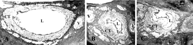
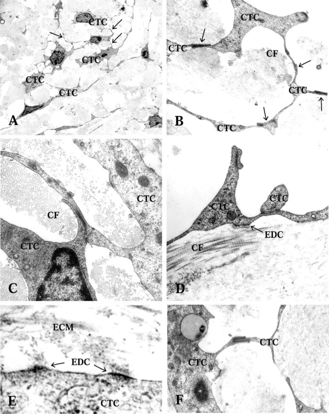
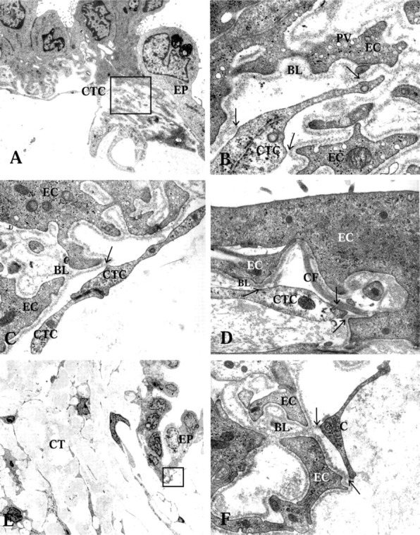
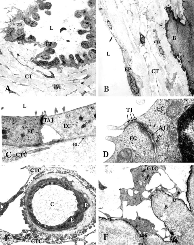
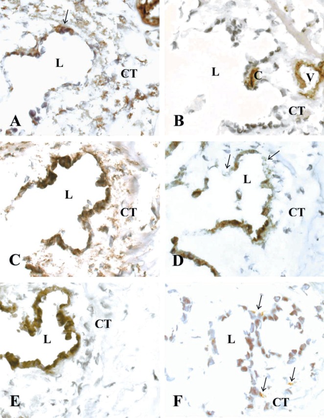
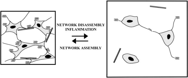
The human endolymphatic duct (ED) and sac of the inner ear have been suggested to control endolymph volume and pressure. However, the physiological mechanisms for these processes remain obscure. We investigated the organization of the periductal interstitial connective tissue cells and extracellular matrix (ECM) in four freshly fixed human EDs by transmission electron microscopy and by immunohistochemistry. The unique surgical material allowed a greatly improved structural and epitopic preservation of tissue. Periductal connective tissue cells formed frequent intercellular contacts and focally occurring electron-dense contacts to ECM structures, creating a complex tissue network. The connective tissue cells also formed contacts with the basal lamina of the ED epithelium and the bone matrix, connecting the ED with the surrounding bone of the vestibular aqueduct. The interstitial connective tissue cells were non-endothelial and non-smooth muscle fibroblastoid cells. We suggest that the ED tissue network forms a functional mechanical entity that takes part in the control of inner ear fluid pressure and endolymph resorption.


Figures

Figure 1
Figure 1
Light microscopy of the human endolymphatic duct (ED) at three different intervals: proximal (A), intermediate (B), and distal (C) ED. The specimen was cut transversely and the light microscopy pictures show the vestibular aqueduct with the ED embedded in loose connective tissue. Note the veins of the vestibular aqueduct and the many bone channels containing blood vessels embedded in loose connective tissue. L, lumen of the ED; CT, connective tissue; B, bone matrix; and V, vein of the vestibular aqueduct.
Figure 2
Figure 2
Transmission electron microscopy of CT cells (CTC), intercellular contacts, and electron-dense contacts (EDC) in the periductal CT. (A,B) CTC intercellular contacts are shown with arrows. Collagen fiber bundles (CF) mostly cut transversly between the CTC network. (C) Two juxtapositioned CTCs with an intercellular contact. (D) Collagen fibers (CF) with typical transmission electron microscopy appearance. Note the electron-dense contact between the ECM and the CTC. (E) Electron-dense contacts between CTC and ECM are seen. (F) A CTC cytoplasmic finger-like indentation is seen, with membrane densifications along the adhesion zone.
Figure 3
Figure 3
Transmission electron microscopy showing connections between the ED epithelium, the CT cells (CTC), and the ECM. (A) Rugose part of the ED epithelium (EP), showing electron-dense and electron-translucent cells and their extensive basal infoldings. Note the CT cell in close vicinity to the basal aspect of the epithelium. Arrows show physical contacts between the CT cells and the basal lamina. Framed area highlighted in B. (B) High-power transmission electron microscopy of CT cell and basal lamina contact spots. Micro-pinocytotic vesicles (PV) are frequent near the cell membrane of the epithelial cells. (C) High-power transmission electron microscopy of a CT cell and epithelial cell contact. Note the CT cells' intercellular contact. (D) The CT cell and epithelial cell contacts are shown with arrows. Note the collagen fiber bundle (CF) just beneath the epithelium in close contact with the basal lamina (BL). (E) Close relationship between the CT cells and the epithelium. Framed area highlighted in F. Arrows showing CT cell and epithelial cell (EC) contacts.
Figure 4
Figure 4
Transmission electron microscopy of the endolymphatic duct (ED) epithelium and the periductal connective tissue (CT). EP, ED epithelium; L, lumen of the ED; CTC, connective tissue cells. (A) Folded epithelium with basal cell processes extending into the surrounding CT, in the intermediate part of the human ED. (B) Flat epithelium with underlying CT in the proximal part of the human ED. Note the interconnecting CT cells of the periductal tissue. B, bone matrix. (C) Flat epithelium with apical microvilli (MV), and intercellular tight and adherens junctions (TAJ). EC, epithelial cell; BL, basal lamina. (D) High-power transmission electron microscopy of intercellular contacts between the epithelial cells. TJ, tight junctions; arrows show three junctional strands; AJ, adherens junctions. (E) A capillary of the periductal CT. E, endothelial cell; P, pericyte; C, lumen of capillary. (F) A CT cell making contacts with the bone matrix (B) and adjacent cell.
Figure 5
Figure 5
Immunohistochemical stainings of the transversely cut human ED with surrounding interstitial CT. L, lumen of the ED; CT, periductal connective tissue; V, vein of the vestibular aqueduct; and C, capillary. Cryostat sections were stained with (A) anti-human vimentin (arrows show positive epithelial cells), (B) anti-human CD31 endothelial cell, (C) anti-human fibroblast (clone 5B5), (D) anti-human fibroblast-specific AS02 (arrow shows negative epithelial cells), (E) anti-pan cytokeratin, and (F) anti-human macrophage CD68 antibodies (arrows show positive cells).
Figure 6
Figure 6
Schematic drawing of a model of the interstitial connective tissue network function and control of tissue swelling. According to this model, the extensive tissue network, formed by CT cells' intercellular contacts and CT cell contacts with the ECM, helps to control interstital fluid pressure by restraining the intrinsic swelling tendency of the ground substance. Inflammation will lead to disorganization of the network structure, probably by affecting the cell-cell and cell-ECM contacts (see Berg et al. 1998) and causing edema to form. Subsequently, the interstitial fluid pressure will increase and lead to impaired endolymph outflow. Eventually endolymphatic hydrops may develop, as seen in patients with Ménière's disease.

References

    1. Altermatt HJ, Gebbers JO, Muller C, Arnold W, Laissue JA. (1990) Human endolymphatic sac: evidence for a role in inner ear immune defence. ORL J Otorhinolaryngol Relat Spec 52:143–148 - PubMed
    1. Bagger-Sjöbäck D, Rask-Andersen H. (1986) The permeability barrier of the endolymphatic sac. A hypothesis of fluid and electrolyte exchange based on freeze fracturing. Am J Otol 7:134–140 - PubMed
    1. Bauwens LJ, De Groot JC, Ramaekers FC, Linthicum F, Veldman JE, Huizing EH. (1991) Differential immunohistochemical detection of cytokeratins and vimentin in the surgically removed human endolymphatic duct and sac. Eur Arch Otorhinolaryngol 248:495–501 - PubMed
    1. Berg A, Ekwall A-KH, Rubin K, Stjernschantz J, Reed RK. (1998) Effect of PGE1, PGI2, and PGF2 alpha analogs on collagen gel compaction in vitro and interstitial pressure in vivo. Am J Physiol 274:H663–671 - PubMed
    1. Chen WT, Singer SJ. (1982) Immunoelectron microscopic studies of the sites of cell-substratum and cell-cell contacts in cultured fibroblasts. J Cell Biol 95:205–222 - PMC - PubMed

Publication types

MeSH terms