. 2003 Nov;51(11):1567-74.
doi: 10.1177/002215540305101116.

Immunocytochemical detection of GLUT2 at the rat intestinal brush-border membrane

Affiliations

Immunocytochemical detection of GLUT2 at the rat intestinal brush-border membrane

Julie A Affleck et al. J Histochem Cytochem. 2003 Nov.

Abstract

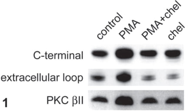
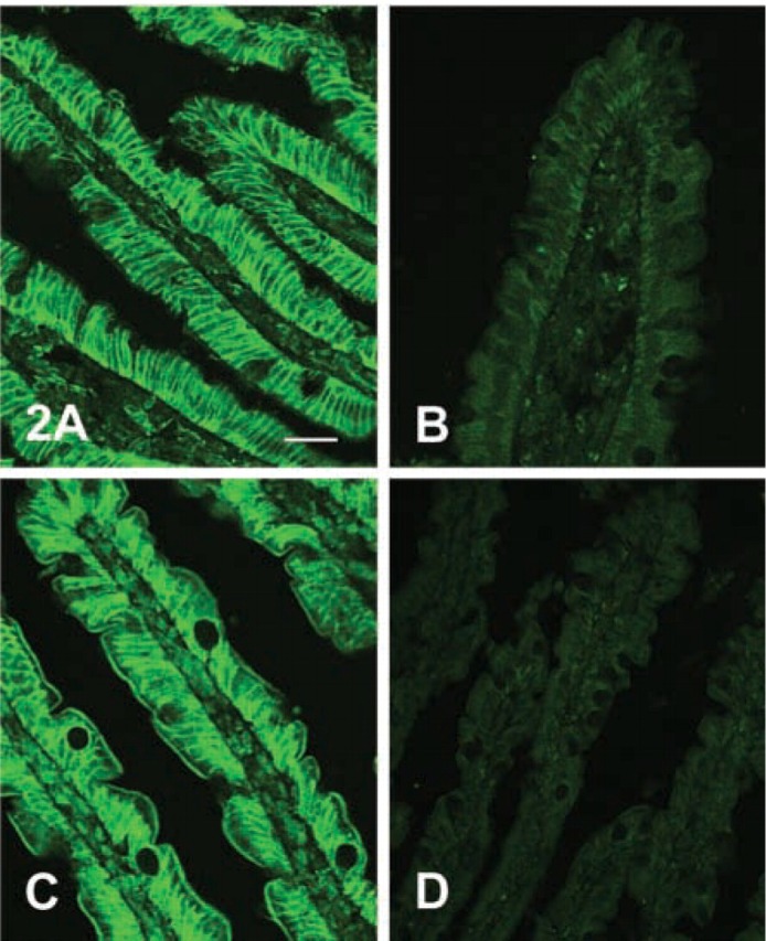
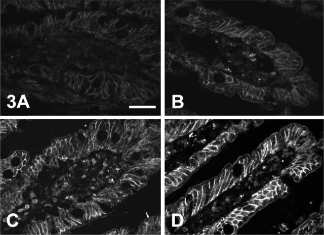
We have proposed a new model of intestinal sugar absorption in which high sugar concentrations promote rapid insertion of the facilitative transporter GLUT2 into the brush-border membrane so that absorptive capacity is precisely regulated to match dietary intake during the assimilation of a meal. However, location of GLUT2 at the brush border by immunocytochemistry has been problematical. We report that control of rapid GLUT2 trafficking and the use of an antibody to a sequence within the large extracellular loop of GLUT2 permits localization of GLUT2 at the brush border. To reveal brush-border GLUT2 fully, it is necessary to digest the sugar chain at the glycosylation site close to the antigenic site. In this way, we have demonstrated by immunocytochemistry PKC-dependent changes in the regulation of brush-border GLUT2 in rat jejunum that correspond to those seen by Western blotting. The functional and immunocytochemical data are now reconciled.


Figures

Figure 1
Figure 1
PMA increases GLUT2 levels at the brush-border membrane of rat jejunum by a PKC-dependent pathway. The jejunum of an anesthetized rat was perfused for 30 min in vivo and then in vitro for 30 min with 5 mM D-fructose, either alone (control) or in the presence of the PKC activator PMA (200 nM). When present, the PKC inhibitor, chelerythrine (chel, 2 μM) was added to the perfusate 15 min before the PMA. After preparation of brush-border membrane vesicles, vesicle protein (15 μg) was separated on 10% SDS-PAGE gels, transblotted onto nitrocellulose, and Western-blotted for GLUT2 using either C-terminal or extracellular loop antisera to rat GLUT2, or a C-terminal monoclonal antibody to PKC βII. For further details see Materials and Methods.
Figure 2
Figure 2
Antiserum to the large extracellular loop but not the C-terminal of GLUT2 detects brush-border membrane GLUT2 in sections of whole jejunum. Rat jejunum was perfused with 5 mM fructose and 200 nM PMA in vitro and then fixed by perfusion with PLP fixative as described in Materials and Methods. Seven-μm sections were then blocked, labeled with GLUT2 antiserum raised in rabbit, and visualized with an FITC-conjugated secondary antibody. Images captured by a confocal microscope were obtained at ×40 magnification: microscope settings for images in A and B were the same. (A) Affinity-purified antibody to residues 508–522 at the C-terminal of rat GLUT2 (1:100 dilution in PBS) and the control with antibody preabsorbed with antigenic peptide (B). (C) Antiserum to residues 40–55 within the large extracellular loop of human GLUT2 obtained from Chemicon International. (1:100 dilution in PBS; C) and the control with antiserum preabsorbed with antigenic peptide (D). Bar = 50 μm.
Figure 3
Figure 3
Digestion of glycosylation sugars reveals GLUT2 at the brush-border membrane. Rat jejunum was perfused with 5 mM fructose in the absence (A,C) and presence (B,D) of 200 nM PMA in vitro and then fixed by perfusion with PLP fixative as described in Materials and Methods. Seven-μm sections were then incubated overnight with PBS alone (A,B) or N-endoglycosidase F (150 U ml−1 in PBS; C,D). Sections were then blocked, labeled with affinity-purified antibody to residues 40–55 within the large extracellular loop of rat GLUT2, and visualized with an FITC-conjugated secondary antibody. Images captured by a confocal microscope were obtained at ×40 magnification. An approximately linear intensity response was obtained by setting the fluorescence saturation limit on the most intense image (D) and the background limit on the least intense image (A). Bar = 50 μm.

References

    1. Au A, Gupta A, Schembri P, Cheeseman CI. (2002) Rapid insertion of GLUT2 into the rat jejunal brush-border membrane promoted by glucagon-like peptide 2. Biochem J 367:247–254 - PMC - PubMed
    1. Brot-Laroche E, Dao MT, Alcade I, Delhomme B, Alvarado F. (1988) Independent modulation by food supply of two distinct sodium-activated D-glucose transport systems in guinea pig jejunal brush-border membrane. Proc Natl Acad Sci USA 85:6370–6373 - PMC - PubMed
    1. Brot-Laroche E, Serrano MA, Delhomme B, Alvarado F. (1986) Temperature sensitivity and substrate specificity of two distinct Na+-activated D-glucose transport systems in guinea pig jejunal brush-border membrane vesicles. J Biol Chem 261:6168–6176 - PubMed
    1. Cheeseman CI. (1993) GLUT2 is the transporter for fructose across the rat intestinal basolateral membrane. Gastroenterology 105:1050–1056 - PubMed
    1. Cheeseman CI, O'Neill D. (1998) Basolateral D-glucose transport activity along the crypt-villus axis in rat jejunum and upregulation induced by gastric inhibitory peptide and glucagon-like peptide-2. Exp Physiol 83:605–616 - PubMed

Publication types