. 2003 Oct 28;100(22):12935-40.
doi: 10.1073/pnas.2231861100. Epub 2003 Oct 17.

The development of diabetes in E2f1/E2f2 mutant mice reveals important roles for bone marrow-derived cells in preventing islet cell loss

Affiliations

The development of diabetes in E2f1/E2f2 mutant mice reveals important roles for bone marrow-derived cells in preventing islet cell loss

Feng X Li et al. Proc Natl Acad Sci U S A. .

Abstract

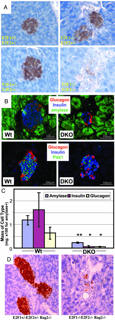
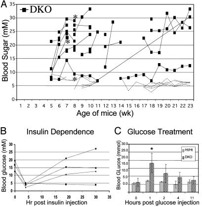
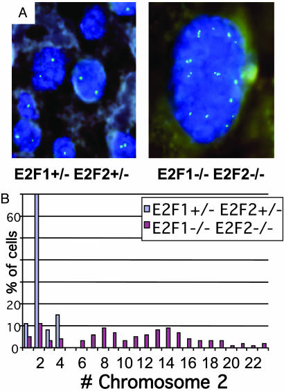
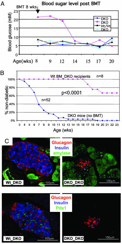
Our studies of mice deficient for the E2F1 and E2F2 transcription factors have revealed essential roles for these proteins in the cell cycle control of pancreatic exocrine cells and the regulation of pancreatic beta cell maintenance. Pancreatic exocrine cells in E2f1-/-E2f2 mutant mice become increasingly polyploid with age, coinciding with severe exocrine atrophy. Furthermore, mice deficient for both E2F1 and E2F2 develop nonautoimmune, insulin-dependent diabetes with high penetrance. Surprisingly, transplantation of wild-type bone marrow can prevent or rescue diabetes in E2f1-/-E2f2-/-mice. We hypothesize that exocrine degeneration results in a destructive environment for beta cells, which can be alleviated by restoration of the hematopoietic system that is also defective in E2f1-/-E2f2-/-mice The demonstration that beta cell maintenance under conditions of stress is influenced by bone marrow-derived cells may provide important insight into the design of therapies to boost islet mass and function in diabetic patients.


Figures

Fig. 2.
Fig. 2.
Age-dependent loss of beta cells in DKO pancreata. Pancreas sections from 2-mo-old (A and D) or 3- to 4-mo-old (B) male mice of the indicated genotypes (note Rag2-/- in D). In A and D, insulin (brown) was detected by immunohistochemistry. In B and C, amylase, insulin, glucagons, and Pdx1 were detected by IF. The same magnification was used within each set. (C) From IF experiments as shown in B (three mice per group), the mass occupied by cells expressing the indicated proteins was determined by morphometric analysis of sections immunostained for insulin, glucagon, and amylase (see Materials and Methods). The percentage of the tissue area occupied by each cell type was multiplied by the total weight of the pancreas to obtain the mass of that cell type per pancreas.
Fig. 1.
Fig. 1.
DKO mice develop nonautoimmune insulin-deficient diabetes. (A) Blood glucose levels of DKO males (black squares) and littermates of other E2f1/2 genotypes (dashed lines with no symbols; two Mt/WT, six Ht/Mt, one WT/Ht, and six Ht/Ht) at different ages. A continuous line represents multiple measurements from a single mouse. Mice designated with an asterisk were also Rag2-/-. (B) Blood glucose levels of four diabetic DKO males at the indicated times after injection of insulin. (C) Blood glucose levels of five nondiabetic DKO females and five Ht/Ht females at the indicated times after injection of glucose. Error bars indicate standard error. *, P < 0.05; **, P < 0.01; ***, P < 0.001 for all figures.
Fig. 3.
Fig. 3.
Exocrine pancreas cells in DKO mice are highly polyploid. (A) FISH analysis of pancreatic exocrine cells from 10-week-old male Ht/Ht and DKO littermates. Chromosome 2-specific signals are in green, and 4′,6-diamidino-2-phenylindole (DAPI) staining of DNA is shown in blue. The same magnifi-cation was used for both pictures (original magnification, ×100). (B) Quantitation of chromosome 2 signals per nuclei from the pancreas sections shown in A. The y axis represents the percentage of cells (of 100 counted) with the indicated number of chromosome 2 signals per nucleus.
Fig. 4.
Fig. 4.
Exocrine degeneration in E2f1/2 mutant mice. (A) Pancreata dissected from mice of the indicated E2f1/E2f2 genotypes were weighed, and pancreas weights are divided into three age groups. Within each age group, statistical significance of differences was determined by comparing to the pancreata weights of Ht/Ht mice. In the second and third age groups, DKO mice also show highly significant pancreata weight decreases compared with Mt/Ht mice (P < 0.001 for each). For the 50-day group, the average ages for Ht/Ht, Mt/Ht, Ht/Mt, and DKO mice were 46.8, 52.3, 51.5, and 47.4 days, respectively. Similarly, the average ages for the 100- and 200-day groups were 162.4, 155.3, 146, and 134.4 days and 270.8, 280.7, 279.8, and 272.3 days, respectively. Age ranges were similar (≈35–75 days, 100–190 days, and 210–405 days) for the three groups, because littermates were generally compared. For Mt/WT and WT/Mt mice in the 200-day group, average ages were 491 (range, 440–570 days) and 463 (range, 440–485 days) days. The average pancreas weight for WT littermates of an average age of 455 days (range, 440–485 days) was 0.23 g. (B) Pancreas sections from a sick 4-mo-old male DKO mouse (Right) and a control littermate (Left) immunostained for insulin (brown). The same magnification was used for both pictures (original magnification, ×20). (C) Pancreas sections from morbid 13-mo-old (Left) and 12-mo-old (Right) male Mt/Ht mice stained with H&E (Left) or immunostained for insulin (Right; dark brown indicates positive cells). Two islets in each picture are indicated with arrows. The same magnification was used for both pictures (original magnification, ×4).
Fig. 5.
Fig. 5.
Prevention or correction of diabetes in DKO mice by BMT. (A) Eight-week-old male mice of the indicated E2f1/E2f2 genotypes were sublethally irradiated and transplanted with WT BM cells (one experiment is shown). Nonfasting blood sugar levels were monitored as shown. (B) Transplantation of WT BM into prediabetic DKO males effectively prevents the development of diabetes. The percentages of DKO that are nondiabetic are plotted as a function of age. DKO males were either untransplanted (blue diamonds) or transplanted with WT BM (pink squares). Two of the DKO recipients of WT BM died at 20 and 26 weeks of unknown causes, but with normal blood sugar levels. Others were healthy at time of death at 16, 18, 23, and 26 weeks. The average age of death (or killing when clearly morbid) of untransplanted DKO males is 15.4 weeks. Five DKO mice received lethal irradiation before BMT, and the other three recipients received sublethal irradiation. Transplanted WT BM was also contained a GFP transgene (7), and >95% of peripheral nucleated blood cells in recipient mice were GFP+ after 1 mo posttransplantation. (C) Anti-insulin, glucagon, amylase, and Pdx1 IF (as indicated) on sections from male DKO recipients of either DKO or WT BM (4 mo old; 2.5 mo after BMT). See Fig. 6D for morphometric quantitations.

References

    1. Kim, S. K. & Hebrok, M. (2001) Genes Dev. 15 111-127. - PubMed
    1. Rane, S. G. & Reddy, E. P. (2000) Front. Biosci. 5 D1-D19. - PubMed
    1. Larsen, S. (1993) Dan. Med. Bull. 40 153-162. - PubMed
    1. Raue, G. & Keim, V. Z. (1999) Gastroenterology 117 Suppl. 1, 4-9. - PubMed
    1. Hardt, P. D., Killinger, A., Nalop, J., Schnell-Kretschmer, H., Zekorn, T. & Klor, H. U. (2002) Pancreatology 2 30-33. - PubMed

Publication types

MeSH terms