. 2003 Oct 28;100(22):12759-64.
doi: 10.1073/pnas.2134254100. Epub 2003 Oct 17.

Extracellular signal-regulated kinase 2 is necessary for mesoderm differentiation

Affiliations

Extracellular signal-regulated kinase 2 is necessary for mesoderm differentiation

Yao Yao et al. Proc Natl Acad Sci U S A. .

Abstract

The extracellular signal-regulated kinase (ERK) is a component of the mitogen-activated protein kinase cascade. Exon 2 of erk2 was deleted by homologous recombination and resulted in embryonic lethality at embryonic day 6.5. erk2 mutant embryos did not form mesoderm and showed increased apoptosis but comparable levels of BrdUrd incorporation, indicating a defect in differentiation. erk2 null embryonic stem (ES) cells exhibited reduced total ERK activity upon serum stimulation, augmented ERK1 phosphorylation, and decreased downstream p90Rsk phosphorylation and activity; yet ES cell proliferation was unaffected. Mutant ES cells were capable of forming mesoderm; however, treatment of mutant ES cells with the mitogen-activated protein kinase kinase inhibitor PD184352 decreased total ERK activity and expression of the mesodermal marker brachyury, suggesting that ERK1 can compensate for ERK2 in vitro. Normal embryos at embryonic day 6.5 expressed activated ERK1/2 in the extraembryonic ectoderm, whereas erk2 mutant embryos had no detectable activated ERK1/2 in this region, suggesting that activated ERK1 was not expressed, and therefore cannot compensate for loss of ERK2 in vivo. These data indicate that ERK2 plays an essential role in mesoderm differentiation during embryonic development.


Figures
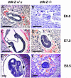
Fig. 1.
Fig. 1.
Histological analysis of normal and presumptive mutant embryos. Hematoxylin and eosin staining of phenotypically normal (A, C, and E) and erk2 presumptive mutant (B, D, and F) embryos between E6.5 and E8.5. The normal embryo elongates and has formed the primitive streak (C, arrow) by E7.5. erk2 presumptive mutants at E7.5 are smaller than normal embryos at E6.5 and E7.5. They increase slightly in size by E7.5 and show no evidence of primitive streak formation (D). At E8.5 normal embryos have turned and cardiac development (arrow) is present (E), whereas presumptive erk2 mutants have not increased in size and appear disorganized (F). Normal embryos have distinguishable neural and cardiac development at E9.5, whereas erk2 presumptive mutants do not increase in size and are resorbed by E9.5 (data not shown). * denotes either + or – for genotype. (Scale bars = 200 μM.)
Fig. 2.
Fig. 2.
Immunohistochemical analysis of normal and mutant embryos. Activated, dp-ERK1/2 immunohistochemistry of E6.5 phenotypically normal (A) and presumptive mutant (B) embryos. Phenotypically normal embryo has strong dp-ERK1/2 staining in a zone of expression in the proximal extraembryonic ectoderm, whereas presumptive mutant embryo does not have this extraembryonic expression. BrdUrd labeling of E7.5 phenotypically normal (C) and presumptive mutant (D) embryos. Both embryos show incorporation of BrdUrd. TUNEL analysis of phenotypically normal (E) and presumptive mutant (F) E7.5 embryos. High levels of TUNEL staining are present in mutant embryo (F) whereas very low levels of TUNEL staining are found in a phenotypically normal embryo (E). Propidium iodide-counterstained phenotypically normal (G) and presumptive mutant (H) embryos at E7.5. * denotes either + or – for genotype. (Scale bars = 200 μM.)
Fig. 3.
Fig. 3.
Analysis of erk2-deficient and WT ES cells. (A) ES cells in response to serum stimulation for 15 min in the absence or presence of the PD184352 MEK inhibitor. Western analysis examining ERK and Rsk levels in WT (TC1, lanes 1–3), heterozygous (365, lanes 4–6), and homozygous mutant (364, lanes 7–9). Cells were serum-starved for 20–24 h before serum stimulation. (B) Kinase activity assays for ERK showing relative kinase activity for serum-starved (lanes 1, 4, and 7), serum-stimulated (lanes 2, 5, and 8), or PD184352-treated (lanes 3, 6, and 9). (C) Kinase activity assays for p90Rsk for serum-starved (lanes 1, 4, and 7), serum-stimulated (lanes 2, 5, and 8), or PD184352-treated (lanes 3, 6, and 9) cells. (D) Growth curves for WT, heterozygous, and homozygous mutant ES lines. ES cells were seeded onto gelatin-coated dishes at a density of 1 × 10⁴ per 60-mm dish. The day after plating is day 0. Cells were counted every day over the next 8 days. Cell numbers were plotted as total cell number compared with days in culture. WT (TC1, dark blue line), heterozygous (365, yellow line), homozygous mutant (364, light blue line), and PD184352-treated WT (pink line).
Fig. 4.
Fig. 4.
In vitro differentiation of erk2-deficient ES cells. (A) Effects of differentiation on Rsk and ERK phosphorylation in ES cells. ES cells were differentiated in CDM in the presence of serum or bFGF and treated with 1 μM or 5 μM PD184352 or vehicle. Lysates from WT TC1 and 364 homozygous mutant ES cells were analyzed by Western blot for p-ERK1/2 and p-Rsk expression. U, undifferentiated, exponentially growing ES cells. p-ERK1/2 and p-Rsk S369 levels decreased upon treatment with PD184352. (B) WT and homozygous mutant ES lines are capable of forming mesoderm in vitro. ES cells were differentiated for 5 days in CDM supplemented with serum and 0, 1, or 5 μM PD184352. RT-PCR analysis for erk2, T (brachyury), and L7 was performed on WT TC1 and mutant 364 ES lines. TC1 ES cells treated with PD184352 have decreased levels of T after 5 days of differentiation in vitro, whereas 364 mutant cells after 5 days of differentiation in the presence of 1 or 5 μM PD184352 have barely detectable levels of T (B Top). Experiments were done in duplicate.

References

    1. Sugiura, N., Suga, T., Ozeki, Y., Mamiya, G. & Takishima, K. (1997) J. Biol. Chem. 272 21575–21581. - PubMed
    1. Lewis, T. S., Shapiro, P. S. & Ahn, N. G. (1998) Adv. Cancer Res. 74 49–139. - PubMed
    1. Lavoie, J. N., Rivard, N., L'Allemain, G. & Pouyssegur, J. (1996) Prog. Cell Cycle Res. 2, 49–58. - PubMed
    1. Whitmarsh, A. J. & Davis, R. J. (2000) Nature 403 255–256. - PubMed
    1. Traverse, S., Gomez, N., Paterson, H., Marshall, C. & Cohen, P. (1992) Biochem. J. 288 351–355. - PMC - PubMed

MeSH terms

Substances

LinkOut - more resources