Clinical Trial
. 2003 Oct;20(2):1400-10.
doi: 10.1016/S1053-8119(03)00391-4.

Putting names to faces: successful encoding of associative memories activates the anterior hippocampal formation

Affiliations
Clinical Trial

Putting names to faces: successful encoding of associative memories activates the anterior hippocampal formation

Reisa Sperling et al. Neuroimage. 2003 Oct.

Abstract

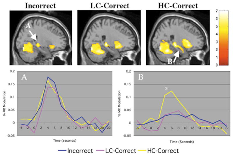
The ability to form associations between previously unrelated items of information, such as names and faces, is an essential aspect of episodic memory function. The neural substrate that determines success vs. failure in learning these associations remains to be elucidated. Using event-related functional MRI during the encoding of novel face-name associations, we found that successfully remembered face-name pairs showed significantly greater activation in the anterior hippocampal formation bilaterally and left inferior prefrontal cortex, compared to pairs that were forgotten. Functional connectivity analyses revealed significant correlated activity between the right and left hippocampus and neocortical regions during successful, but not attempted, encoding. These findings suggest that anterior regions of the hippocampal formation, in particular, are crucial for successful associative encoding and that the degree of coordination between hippocampal and neocortical activity may predict the likelihood of subsequent memory.


Figures

Fig. 1
Fig. 1
Successful vs. Failed Encoding. Statistical activation maps demonstrating greater response during encoding of Face-Name pairs that were subsequently successfully remembered (High Confidence Correct) compared to Face-Name Pairs that were forgotten (Incorrect). Activation within the hippocampal formation is shown in three image planes: coronal (top), sagittal (middle), and transaxial (bottom), and was located in the anterior hippocampus bilaterally, and the right entorhinal cortex. Analyses performed with SPM99, and statistical activation maps (color bar represents T scores) of the event-related data averaged across subjects are displayed on an anatomic T1 image template.
Fig. 2
Fig. 2
Attempted Encoding vs. Fixation. Activation maps for each of the attempted encoding conditions compared to visual fixation (color bar represents T scores): Face-Name pairs that were subsequently forgotten (Incorrect), Remembered with Low Confidence (LC-Correct), and Remembered with High Confidence (HC-Correct). Within the hippocampal formation, Incorrect (top left) and LC-Correct (top middle) demonstrated significant activation in mid-posterior hippocampus only, while HC-Correct (top right) demonstrated activation along the entire longitudinal axis of the hippocampus, including the anterior hippocampus. MR signal time courses were extracted for a three-dimensional region of interest (4-mm radius) for the posterior hippocampus (bottom left, A: centered at 21, −30, −3) and the anterior hippocampus (bottom right, B: centered at 24, −12, −15). The percent MR signal modulation for each condition is indicated on the y-axis and time in seconds is indicated on the x-axis. All encoding conditions (HC-Correct, LC-Correct, and Incorrect) showed similar response in mid- to posterior hippocampus, while HC-Correct responses were significantly higher (P < 0.00001) than LC-Correct and Incorrect in the anterior hippocampus.
Fig. 3
Fig. 3
Analysis of confidence and accuracy. Activation maps for HC-Correct vs. LC-Correct (yellow) and HC-Incorrect vs. LC-Incorrect (red) are displayed on coronal slices: prefrontal regions shown on left at y = 27, and hippocampal regions shown on the right at y = −12). No significant overlap was found using a conjunction analysis at the threshold of P < 0.001, but at the lower threshold atP < 0.01 (shown in figure), the only region showing overlap between confidence and accuracy was found in the left inferior prefrontal cortex. There was bilateral activation in the anterior hippocampal formation for the HC-correct vs. LC-correct contrast, but no activation within the hippocampal formation for the HC-Incorrect vs. LC-Incorrect even at the lower threshold of P < 0.01.

References

    1. Barbas H, Blatt GJ. Topographically specific hippocampal projections target functionally distinct prefrontal areas in the rhesus monkey. Hippocampus. 1995;5:511–533. - PubMed
    1. Brewer JB, Zhao Z, Desmond JE, Glover GH, Gabrieli JD. Making memories: brain activity that predicts how well visual experience will be remembered. Science. 1998;281:1185–1187. - PubMed
    1. Buckner RL, Wheeler ME, Sheridan MA. Encoding processes during retrieval tasks. J Cogn Neurosci. 2001;13:406–415. - PubMed
    1. Bunsey M, Eichenbaum H. Selective damage to the hippocampal region blocks long-term retention of a natural and nonspatial stimulus-stimulus association. Hippocampus. 1995;5:546–556. - PubMed
    1. Cabeza R, Dolcos F, Graham R, Nyberg L. Similarities and differences in the neural correlates of episodic memory retrieval and working memory. NeuroImage. 2002;16:317–330. - PubMed

Publication types