. 2003 Oct 20;198(8):1213-24.
doi: 10.1084/jem.20030918.

Elimination in vivo of developing T cells by natural killer cells

Affiliations

Elimination in vivo of developing T cells by natural killer cells

Eckart Schott et al. J Exp Med. .

Abstract

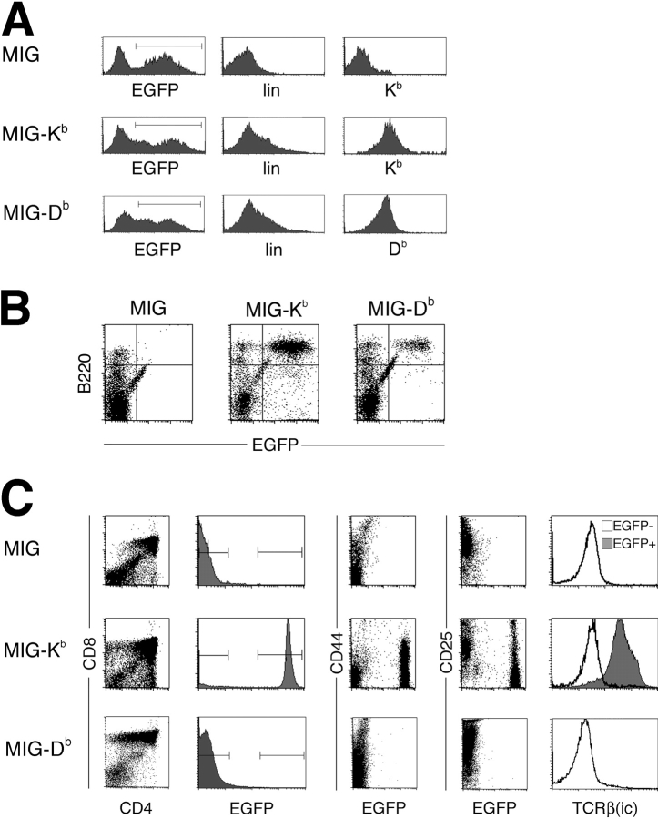
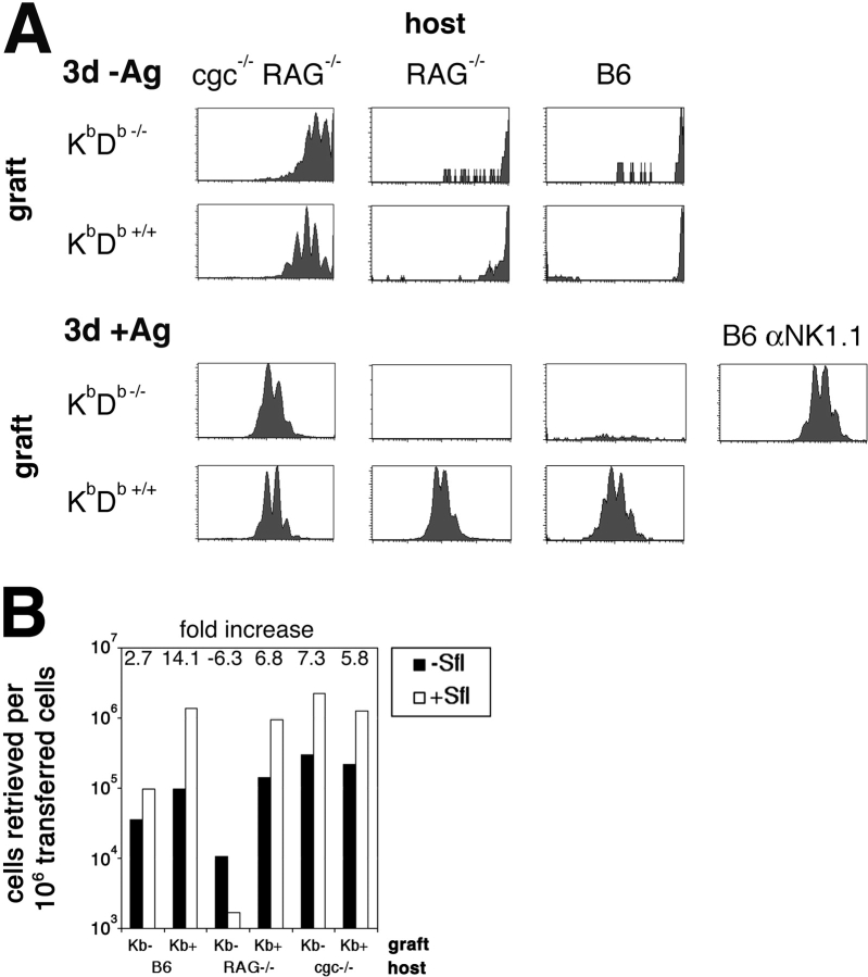
Natural killer cells gauge the absence of self class I MHC on susceptible target cells by means of inhibitory receptors such as members of the Ly49 family. To initiate killing by natural killer cells, a lack of inhibitory signals must be accompanied by the presence of activating ligands on the target cell. Although natural killer cell-mediated rejection of class I MHC-deficient bone marrow (BM) grafts is a matter of record, little is known about the targeting in vivo of specific cellular subsets by natural killer cells. We show here that development of class I MHC-negative thymocytes is delayed as a result of natural killer cell toxicity after grafting of a class I MHC-positive host with class I MHC-negative BM. Double positive thymocytes that persist in the presence of natural killer cells display an unusual T cell receptor-deficient phenotype, yet nevertheless give rise to single positive thymocytes and yield mature class I MHC-deficient lymphocytes that accumulate in the class I MHC-positive host. The resulting class I MHC-deficient CD8 T cells are functional and upon activation remain susceptible to natural killer cell toxicity in vivo. Reconstitution of class I MHC-deficient BM precursors with H2-K(b) by retroviral transduction fully restores normal thymic development.


Figures

Figure 1.
Figure 1.
Generation of DP and SP thymocytes from KbDb−/− BM grafts is delayed. (A) BM from KbDb−/− and KbDb+/+ mice was stained with lineage marker antibodies (lin) and with antibodies against c-kit and sca-1 to establish the frequency of progenitor cells. (B) BM from KbDb−/− or KbDb+/+ mice was transferred into sublethally irradiated RAG−/− hosts. Mice were killed at the times indicated, and thymi were analyzed by flow cytometry after staining for CD4, CD8, and intracellular TCRβ. (C) The total number of thymocytes retrieved at different time points after transfer of KbDb−/− or KbDb+/+ BM is indicated. (D) Spleen cells from chimeras that had received KbDb−/− or KbDb+/+ BM were analyzed by flow cytometry 10 wk after BM transfer. (E) BM from KbDb−/−, β2m−/−, TAP−/−, or KbDb+/+ (B6) mice was transferred into sublethally irradiated RAG−/− hosts. Thymi were analyzed at 4 wk by flow cytometry after staining for CD4 and CD8.
Figure 2.
Figure 2.
The delay in development of KbDb−/− thymocytes is due to NK cell toxicity. (A) BM from KbDb−/− mice was transferred into sublethally irradiated RAG−/− hosts that were treated with NK1.1 antibody or the appropriate isotype control. Mice were killed at the times indicated, and thymi were analyzed by flow cytometry after surface staining for CD4, CD8, and intracellular TCRβ. (B) BM from OT-I KbDb−/− or OT-I KbDb+/+ mice was transferred into sublethally irradiated perforin−/− hosts. Mice were killed at the times indicated, and thymi were analyzed by flow cytometry after surface staining for CD4 and CD8. Since endogenous thymocytes are present, the presence of thymocytes expressing the transgenic TCR OT-I was visualized by staining with Kb(SIINFEKL) tetramer. (C) BM from KbDb−/− or KbDb+/+ mice was transferred into sublethally irradiated RAG−/− hosts. Mice were killed at 4 wk, and thymi and spleens were analyzed by flow cytometry after surface staining for CD3 and NK1.1, and intracellular staining for IFNγ. (D) Experimental setup as in C, but staining was performed with anti-CD4, anti-CD8, and anti-fas antibodies. Data shown in the histograms result from gating on DP thymocytes.
Figure 3.
Figure 3.
KbDb−/− DP thymocytes developing in the presence of NK cells display an immature phenotype. (A) BM from KbDb−/− or KbDb+/+ mice was transferred into sublethally irradiated RAG−/− hosts. Mice were killed at 4 wk, and thymi were analyzed by flow cytometry after surface staining for CD4, CD8, TCRβ, CD25, and intracellular staining for TCRβ. Data shown in the histograms results from gating on DP thymocytes as shown in the left panels. (B) BM from OT-I KbDb−/− or OT-I KbDb+/+ mice was transferred into sublethally irradiated RAG−/− hosts. Mice were killed at the times indicated, and thymi were analyzed by flow cytometry after surface staining for CD4, CD8, and with Kb(SIINFEKL) tetramer to visualize presence of the OT-I TCR. (C) Experimental setup as in A, but staining was performed with anti-CD4, anti-CD8, and anti-H2-Kb antibodies in chimeras 8 wk after the transfer of BM. Histograms show data resulting from gating on DN, DP, or CD8 SP thymocytes.
Figure 4.
Figure 4.
KbDb−/− thymocytes that persist in the presence of NK cells show reduced proliferation. BM from KbDb−/− or KbDb+/+ mice was transferred into sublethally irradiated RAG−/− hosts. BrdU labeling was performed at 4 wk by two i.p. injections at 0 and 4 h. Mice were killed 24 h later, and thymi were analyzed by flow cytometry after surface staining for CD4, CD8, and intracellular staining for BrdU. Data shown in the histograms results from gating on DP or DN thymocytes as indicated.
Figure 5.
Figure 5.
Phenotypically distinct KbDb−/− thymocytes generated in the presence on NK cells give rise to polyclonal mature T cells. (A) BM from KbDb−/− or KbDb+/+ mice was transferred into sublethally irradiated RAG−/− hosts. Mice were killed at 10 wk, and spleens were analyzed by flow cytometry after surface staining with anti-CD4 and anti-CD8 antibodies and with a panel of different anti-Vβ antibodies. The percentage of spleen cells staining with each individual anti-Vβ antibody is shown for animals grafted with KbDb−/− or KbDb+/+ BM. (B) BM from KbDb−/− or KbDb+/+ mice was transferred into sublethally irradiated RAG−/− hosts. Mice were killed at 3 wk, and one thymic lobe was analyzed by flow cytometry after surface staining for CD4 and CD8. The corresponding thymic lobe was transplanted under the kidney capsule of RAG−/− cγc−/− mice. Recipient mice were killed 5 wk after the procedure, and thymic grafts and spleens were analyzed by flow cytometry after staining with anti-CD4, anti-CD8, anti-Vα2, and anti-Vβ5 antibodies.
Figure 6.
Figure 6.
Reconstitution of KbDb−/− BM with H2-Kb restores normal thymocyte development. (A) cDNAs for H2-Kb and H2-Db were generated by RT-PCR and subcloned into MIG-R. BM was harvested from KbDb−/− mice and infected in vitro with retrovirus encoding for EGFP and H2-Kb (MIG-Kb), EGFP and H2-Db (MIG-Db), or EGFP alone (MIG). Flow cytometry analysis of cultured cells was performed at day 5 of culture after staining with lineage marker antibodies and anti–H2-Kb or anti–H2-Db antibodies. Data shown in the right two histograms results from gating on EGFP+ cells as shown in the left histogram. (B) Equal numbers of EGFP+ lin BM cells infected with the indicated virus in vitro were transferred into sublethally irradiated RAG−/− hosts. Recipient mice were killed at 4 wk, and spleens were analyzed by flow cytometry after staining for B220. (C) Equal numbers of EGFP+ lin BM cells infected with the indicated virus in vitro were transferred into sublethally irradiated RAG−/− hosts. Recipient mice were killed at 4 wk, and thymi were analyzed by flow cytometry after staining for CD4, CD8, CD44, CD25, and intracellular staining for TCRβ. Data is representative for two independent experiments with a total of six chimeras for each group.
Figure 7.
Figure 7.
Mature CD8 T cells generated from KbDb−/− grafts in the presence of NK cells remain susceptible to NK cell killing. (A) BM from OT-I KbDb−/− or OT-I KbDb+/+ mice was transferred into sublethally irradiated RAG−/− hosts. LN cells were harvested 10 wk after the transfer, enriched for CD8 T cells, labeled with CFSE, and transferred in equal numbers into recipient mice as indicated. Some mice were challenged with the antigenic peptide SIINFEKL i.p. 24 h after the transfer (+Ag). Mice were killed 72 h after the transfer, and spleen and LN (in the case of RAG−/− and B6 mice) were harvested. Cells were stained with anti-CD8 antibody and with Kb(SIINFEKL) tetramer to detect transgenic CD8 T cells. CFSE dilution profiles are shown after gating on CD8+ tetramer+ cells. (B) Experimental setup as in A. The relative number of OT-I T cells retrieved from recipient animals is shown for mice grafted with KbDb−/− and KbDb+/+ BM and the effect of challenge with the antigenic peptide (SH) on the number of OT-I cells is indicated above each pair of bars. Data are presented as mean values from duplicate experiments.

Similar articles

Cited by

References

    1. Yu, Y.Y., T. George, J.R. Dorfman, J. Roland, V. Kumar, and M. Bennett. 1996. The role of Ly49A and 5E6(Ly49C) molecules in hybrid resistance mediated by murine natural killer cells against normal T cell blasts. Immunity. 4:67–76. - PubMed
    1. Bix, M., N.S. Liao, M. Zijlstra, J. Loring, R. Jaenisch, and D. Raulet. 1991. Rejection of class I MHC-deficient haemopoietic cells by irradiated MHC-matched mice. Nature. 349:329–331. - PubMed
    1. Lanier, L.L. 1998. NK cell receptors. Annu. Rev. Immunol. 16:359–393. - PubMed
    1. Yokoyama, W.M. 1995. Natural killer cell receptors. Curr. Opin. Immunol. 7:110–120. - PubMed
    1. Yokoyama, W.M. 1993. Recognition structures on natural killer cells. Curr. Opin. Immunol. 5:67–73. - PubMed

Publication types

Substances