. 2003 Oct 28;100(22):13048-53.
doi: 10.1073/pnas.1735415100. Epub 2003 Oct 20.

Expression of T cell receptor beta locus in central nervous system neurons

Affiliations

Expression of T cell receptor beta locus in central nervous system neurons

Josh Syken et al. Proc Natl Acad Sci U S A. .

Abstract

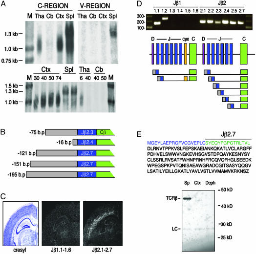
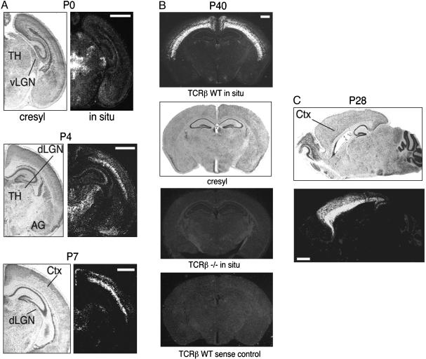
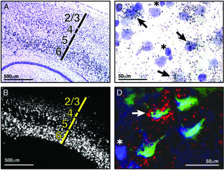
MHC class I proteins are cell-surface ligands that bind to T cell receptors and other immunoreceptors and act to regulate the activation state of immune cells. Recent work has shown that MHC class I genes and CD3zeta, an obligate component of T cell receptors, are expressed in neurons, are regulated by neuronal activity, and function in neuronal development and plasticity. A search for additional neuronally expressed T cell receptor components has revealed that the T cell antigen receptor beta (TCRbeta) locus is expressed in neurons of the murine central nervous system and that this expression is dynamically regulated over development. In neonates, expression is most abundant in various thalamic nuclei. At later ages and in adults, thalamic expression fades and cortical expression is robust, particularly in layer 6. In T cells, protein-encoding transcripts are produced only after recombination of the TCRbeta genomic locus, which joins variable, diversity, and joining regions, a process that creates much of the diversity of the immune system. We detect no genomic recombination in neurons. Rather, transcripts begin in regions upstream of several joining regions, and are spliced to constant region segments. One of the transcripts encodes a hypothetical 207-aa, 23-kDa protein, which includes the TCRbeta J2.7 region, and the entire C region. These observations suggest that TCRbeta may function in neurons.


Figures

Fig. 3.
Fig. 3.
Neuronal TCRβ is transcribed from unrearranged genomic loci. (A Top) Northern blot analysis of RNA derived from P9 thalamus (Tha), P29 cerebellum (Cb), P30 cortex (Ctx), and adult spleen (Spl), probed with TCRβ C region or mix of V region probes. (Bottom) RNA derived from P30, P40, P50, and P74 cortex, adult spleen, P6 and P40 thalamus, and P40 and P50 cerebellum, probed for TCRβ C region. M, molecular weight markers. (B) RACE-derived sequences indicate that the 5′ ends of neuronal TCRβ are identical with genomic sequences 5′ to several of the TCRβ J regions. Numbers indicate bases from the beginning of transcript to Jβ exon junction. (C) In situ hybridization of adult mouse brain sections by using probes for genomic sequence spanning Jβ1.1–1.6 or Jβ2.1–2.7 indicates that the majority of neuronal TCRβ mRNA includes Jβ2 regions. (D) RT-PCR by using primers ≈20 bp 5′ to the start of each Jβ coding region indicated, plus a common C region 3′ primer, indicate that neuronal TCRβ transcripts include at least 8 of the 12 Jβ regions and include noncoding intergenic sequences upstream of Jβ regions. One sequence that included Jβ1.2 also included the Cβ0 exon. (E) Hypothetical truncated TCRβ protein sequence. (Top) Amino acid sequence of hypothetical protein encoded by genomic region 5′ to Jβ2.7 (blue), Jβ2.7 (green), and C region (black). (Bottom) Immunoprecipitation/Western blot analysis of saline-perfused brain tissue using an antibody to the C region of TCRβ. LC, light chain; Sp, spleen; Ctx, cortex; Dcph, diencephalon. Analyzed were 100 μg of spleen tissue and 5 mg of cortex and diencephalon.
Fig. 1.
Fig. 1.
TCRβ mRNA is expressed in the mouse brain. Cryostat sections from postnatal days 0, 4, 7, 28, or 40 were either stained with cresyl violet or hybridized with a probe for TCRβ C regions. (A) Coronal sections. (B) P40 TCRβ-/- and TCRβ wild-type sense control sections are similar to the P40 section hybridized with TCRβ antisense probe. (C) Sagittal sections, anterior to the left. TH, thalamus; AG, amygdala; dLGN, dorsal lateral geniculate nucleus; vLGN, ventral lateral geniculate nucleus; CTX, cortex. (Scale bars, 1 mm.)
Fig. 2.
Fig. 2.
TCRβ is expressed in a subset of cortical neurons. (A and B) P40 coronal section showing cortical layers 1–6 (A) and TCRβ C region in situ hybridization shown in darkfield optics (B) (silver grains appear white). (C) High-power image of layer 6 cortical cells double-stained with cresyl violet (purple) and TCRβ in situ hybridization (black silver grains). Arrows point to large pale nuclei with associated silver grains; asterisks indicate smaller dark nuclei. (D) Triple labeling of cortical layer 6 neurons from posterior cortical region. Hoechst-stained nuclei (blue), retrograde label from Fluoro-Gold injection in striatum (green), and TCRβ in situ hybridization (red) are shown. Arrow indicates triple-labeled cell; asterisk indicates nucleus of a cell with no retrograde label and no TCRβ expression.

References

    1. Ploegh, H. L., Orr, H. T. & Strominger, J. L. (1981) Cell 24 287-299. - PubMed
    1. Rammensee, H. G., Falk, K. & Rotzschke, O. (1993) Curr. Opin. Immunol. 5 35-44. - PubMed
    1. Lampson, L. A. (1987) Trends Neurosci. 10 211-216.
    1. Wong, G. H., Bartlett, P. F., Clark-Lewis, I., Battye, F. & Schrader, J. W. (1984) Nature 310 688-691. - PubMed
    1. Wong, G. H., Bartlett, P. F., Clark-Lewis, I., McKimm-Breschkin, J. L. & Schrader, J. W. (1985) J. Neuroimmunol. 7 255-278. - PubMed

Publication types

MeSH terms

Substances