. 2003 Oct 28;100(22):12989-94.
doi: 10.1073/pnas.2134250100. Epub 2003 Oct 20.

Genetic requirements for mycobacterial survival during infection

Affiliations

Genetic requirements for mycobacterial survival during infection

Christopher M Sassetti et al. Proc Natl Acad Sci U S A. .

Abstract

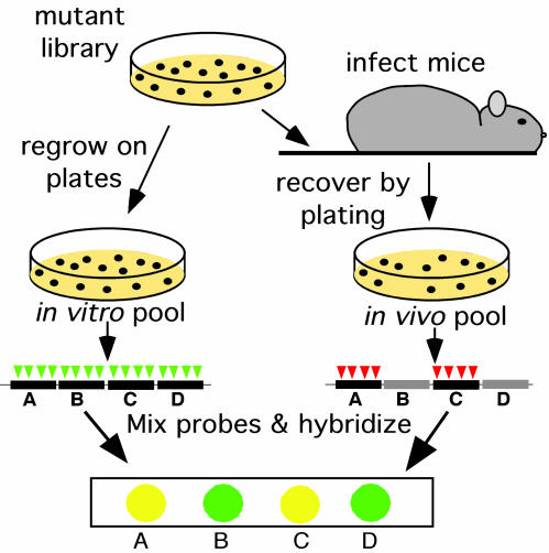
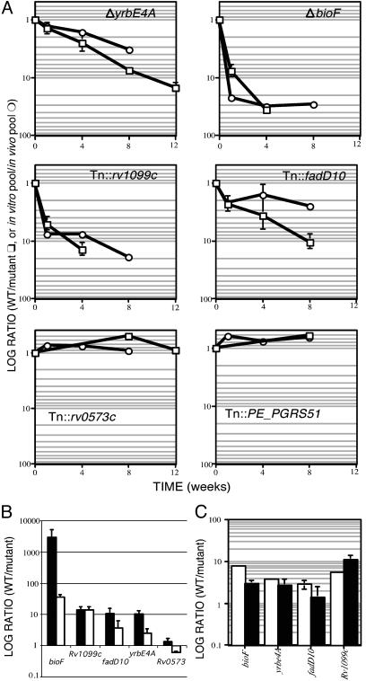
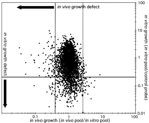
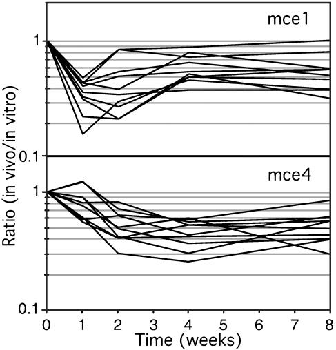
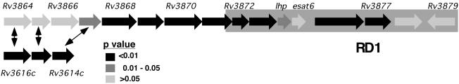
Despite the importance of tuberculosis as a public health problem, we know relatively little about the molecular mechanisms used by the causative organism, Mycobacterium tuberculosis, to persist in the host. To define these mechanisms, we have mutated virtually every nonessential gene of M. tuberculosis and determined the effect disrupting each gene on the growth rate of this pathogen during infection. A total of 194 genes that are specifically required for mycobacterial growth in vivo were identified. The behavior of these mutants provides a detailed view of the changing environment that the bacterium encounters as infection proceeds. A surprisingly large fraction of these genes are unique to mycobacteria and closely related species, indicating that many of the strategies used by this unusual group of organisms are fundamentally different from other pathogens


Figures

Fig. 1.
Fig. 1.
TraSH to identify attenuated mutants. C57BL/6J mice were infected intravenously with 106 colony-forming units of the mutant library. At the indicated times after infection, surviving bacteria were recovered by plating on agar medium (in vivo pools). The in vitro pool was generated by replating the library. The in vitro pool contained mutants with insertions (represented by triangles) in each nonessential gene (black bars). Mutants harboring insertions in genes that are specifically required for survival in the mouse spleen (gray bars) were lost from the in vivo pool. Genomic DNA was isolated from each pool, and TraSH probe was generated that was complementary to the chromosomal sequence flanking each insertion in the pool (4). The probes from the two pools were labeled with different colored fluorophores and mixed. Probes were then hybridized to a microarray onto which DNA fragments (features) were immobilized that were complementary to each gene in the genome of M. tuberculosis. Features that hybridized to the probe from the in vitro, but not the in vivo, pool represented genes that were specifically required for growth in the mouse.
Fig. 2.
Fig. 2.
TraSH accurately predicts the in vivo growth characteristics of individual mutants. (A) Average TraSH ratios for the indicated genes (in vitro pool/in vivo pool) are plotted on a log scale as a function of time (circles). Individual strains carrying mutations in these genes were mixed with wild-type bacteria and inoculated into mice. The ratio (wild type/mutant) at each time point after infection is plotted (squares) on the same scale as the TraSH data. Data were normalized so the ratio of the inoculum equaled 1. Error bars represent standard deviations. (B) Growth of individual mutants in lung (filled bars) and spleen (open bars). Data were collected as in A from organs harvested after 4 weeks of infection except for Tn::Rv0573 (8 weeks). (C) Attenuated strains do not have in vitro growth defects. Each mutant strain was mixed with wild-type H37Rv and grown in triplicate broth cultures. The ratio of wild type to mutant was determined after inoculation (open bars) and after 10 days of growth (filled bars). Only the ratio for the ΔbioF mutant changed significantly (P = 0.0005; mutant grew faster than wild type).
Fig. 3.
Fig. 3.
Comparison of in vivo and in vitro growth rates. Predicted in vivo growth rates are represented as TraSH ratios (8-week in vivo pool/in vitro pool) for each gene on the x axis. Solid vertical line represents cutoff value of 0.4×, which defines attenuating mutations. Dotted line indicates cutoff for the definition of mutants with increased in vivo growth rates. Relative in vitro growth rates were determined previously (4) and are represented on the y axis as the ratio of TraSH probe from the in vitro pool divided by a control probe of labeled chromosomal DNA. Horizontal line represents 0.2× cutoff value, identifying mutants with in vitro growth defects.
Fig. 4.
Fig. 4.
In vivo behavior of mce mutants. TraSH ratios for each gene in the mce1 (Rv0169-Rv0178) or mce4 (Rv3492c-Rv3501c) loci are plotted as a function of time (yrbE1A and yrbE1B of mce1 locus were omitted because of lack of data). TraSH data compare mutants grown in vitro with those isolated from mouse spleens.
Fig. 5.
Fig. 5.
Genes surrounding esat6 are required for in vivo survival. The esat-6 locus is depicted and shaded according to the significance level (P value) that the ratio for each gene differs from 1 (at the 4-week time point). Homology between Rv3614c-Rv3616c and the esat-6 locus is indicated by arrows. “RD1” refers to the region deleted in M. bovis bacillus Calmette–Guérin.

References

    1. Flynn, J. L. & Chan, J. (2001) Annu. Rev. Immunol. 19, 93-129. - PubMed
    1. Glickman, M. S. & Jacobs, W. R., Jr. (2001) Cell 104, 477-485. - PubMed
    1. Sassetti, C. M., Boyd, D. H. & Rubin, E. J. (2001) Proc. Natl. Acad. Sci. USA 98, 12712-12717. - PMC - PubMed
    1. Sassetti, C. M., Boyd, D. H. & Rubin, E. J. (2002) Mol. Microbiol. 48, 77-84. - PubMed
    1. Bardarov, S., Bardarov, S., Jr., Pavelka, M. S., Jr., Sambandamurthy, V., Larsen, M., Tufariello, J., Chan, J., Hatfull, G. & Jacobs, W. R., Jr. (2002) Microbiology 148, 3007-3017. - PubMed

Publication types

Substances

LinkOut - more resources