. 2003 Oct 28;100(22):12618-23.
doi: 10.1073/pnas.2134510100. Epub 2003 Oct 20.

Active sites of thioredoxin reductases: why selenoproteins?

Affiliations

Active sites of thioredoxin reductases: why selenoproteins?

Stephan Gromer et al. Proc Natl Acad Sci U S A. .

Abstract

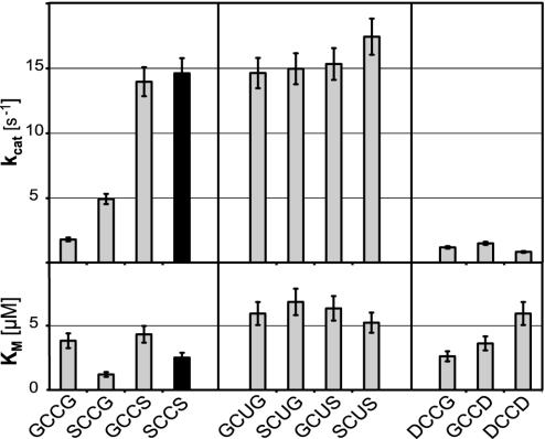
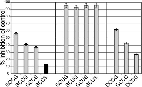
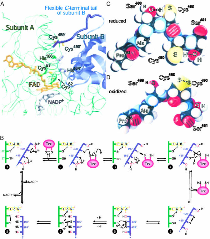
Selenium, an essential trace element for mammals, is incorporated into a selected class of selenoproteins as selenocysteine. All known isoenzymes of mammalian thioredoxin (Trx) reductases (TrxRs) employ selenium in the C-terminal redox center -Gly-Cys-Sec-Gly-COOH for reduction of Trx and other substrates, whereas the corresponding sequence in Drosophila melanogaster TrxR is -Ser-Cys-Cys-Ser-COOH. Surprisingly, the catalytic competence of these orthologous enzymes is similar, whereas direct Sec-to-Cys substitution of mammalian TrxR, or other selenoenzymes, yields almost inactive enzyme. TrxRs are therefore ideal for studying the biology of selenocysteine by comparative enzymology. Here we show that the serine residues flanking the C-terminal Cys residues of Drosophila TrxRs are responsible for activating the cysteines to match the catalytic efficiency of a selenocysteine-cysteine pair as in mammalian TrxR, obviating the need for selenium. This finding suggests that the occurrence of selenoenzymes, which implies that the organism is selenium-dependent, is not necessarily associated with improved enzyme efficiency. Our data suggest that the selective advantage of selenoenzymes is a broader range of substrates and a broader range of microenvironmental conditions in which enzyme activity is possible.


Figures

Fig. 1.
Fig. 1.
Comparison of kinetic parameters of wild-type and mutant DmTrxR. The kcat (Upper) and Km (Lower) values shown here were determined by using DmTrx-2 as a substrate (GHOST assay). The C-terminal tetrapeptide sequences of the respective enzyme are indicated on the x axis. The results for the wild-type enzyme represented by SCCS are highlighted in black. The kcat values shown for the selenoenzymes were normalized for the presence of truncated, inactive enzyme. Note that the wild-type enzyme's (SCCS) activity does not differ significantly from the kcat values of the Sec mutants.
Fig. 2.
Fig. 2.
Half-reactions of wild-type enzyme and mutant forms. Each protein was reduced with sodium borohydride. Reduced protein was mixed in the stopped-flow spectrophotometer with various concentrations of oxidized DmTrx. Solid lines represent the reduced enzyme species with no Trx added. Dotted spectra depict the enzyme species after addition of two equivalents of oxidized Trx. In the SCCG case, the dashed line represents three equivalents of Trx, and the dash-dotted line represents five equivalents. For clarity, the spectra of GCCG are shifted by 0.05 units. The absorbance of the GCCS mutant was essentially identical to that of GCCG, whereas the SCCS mutant had essentially the same absorbance as SCCG (data not shown). (Inset) The reductive half-reaction of wild-type enzyme (SCCS) compared with three mutant forms (GCCG, SCCG, and GCCS). The protein was mixed in a stopped-flow spectrophotometer with four equivalents of NADPH per subunit, and the change in absorbance at 462 nm, representing the redox state of the flavin, was recorded over time. All enzymes were used at similar concentrations (≈18 μM subunits). The absorption starting point (0.2) was shifted for the GCCS, SCCG, and GCCG mutants for clarity. Note that the time axis is in logarithmic scale. See text for details.
Fig. 3.
Fig. 3.
Inhibition by auranofin. Auranofin, an effective inhibitor of human TrxRs, was analyzed for its inhibitory effect on wild-type DmTrxR (highlighted in black) and the DmTrxR mutants. Shown is the percentage of inhibition compared with an auranofin-free control. In all cases, 1 μM auranofin was used and the assays were carried out as in Fig. 1.
Fig. 4.
Fig. 4.
(A) Computer model of DmTrxR. The protein backbones of the two subunits in the dimeric enzyme are shown as thin green strands (subunit A) and blue ribbons (subunit B). The flexible C-terminal tail of subunit B is shown as a thick blue strand. The FAD (orange) and nicotinamide (gray) of NADP+, bound to subunit A, are represented as stick models. The side chains of the N-terminal and C-terminal redox-active site (Cys57 and Cys62 and Cys489′ and Cys490′, respectively), as well as the putative base catalysts, His106 and His464′, are also shown as stick models, with the sulfurs in yellow and the nitrogens of His in blue. The van der Waals radii of these amino acids are dotted. This model is based on the crystal structure of rat TrxR U498C (21) (Protein Data Bank ID code 1H6V) by using raswin v2.7.2 for visualization (www.bernstein-plus-sons.com/software/RasMol_2.7.2/doc/rasmol.html). (B) A proposed mechanism for DmTrxR. The dotted arrows between the thiolate of Cys-62 and the flavin indicate charge–transfer interactions. The change in conformation is indicated by the juxtaposition of the two polypeptide chains. See text for other details. (C) Corey–Pauling–Koltun space-filling model of the C terminus of wild-type-DmTrxR in the dithiol state. (D) Corey-Pauling-Koltun space-filling model of the C terminus in the disulfide state. All peptide bonds shown in C and D are trans-configured.

References

    1. Bösl, M. R., Takaku, K., Oshima, M., Nishimura, S. & Taketo, M. M. (1997) Proc. Natl. Acad. Sci. USA 94 5531–5534. - PMC - PubMed
    1. Böck, A., Forchhammer, K., Heider, J. & Baron, C. (1991) Trends Biochem. Sci. 16 463–467. - PubMed
    1. Gromer, S., Urig, S. & Becker, K. (2003) Med. Res. Rev. 24 in press. - PubMed
    1. Gladyshev, V. N., Jeang, K. T. & Stadtman, T. C. (1996) Proc. Natl. Acad. Sci. USA 93 6146–6151. - PMC - PubMed
    1. Tamura, T. & Stadtman, T. C. (1996) Proc. Natl. Acad. Sci. USA 93 1006–1011. - PMC - PubMed

Publication types

MeSH terms

LinkOut - more resources