

Regulation of amylin release from cultured rabbit gastric fundic mucosal cells
- PMID: 14572315
- PMCID: PMC269984
- DOI: 10.1186/1472-6793-3-13
Regulation of amylin release from cultured rabbit gastric fundic mucosal cells
Abstract
Background: Amylin (islet amyloid polypeptide) is a hormone with suggested roles in the regulation of glucose homeostasis, gastric motor and secretory function and gastroprotection. In the gastric mucosa amylin is found co-localised with somatostatin in D-cells. The factors regulating gastric amylin release are unknown. In this study we have investigated the regulation of amylin release from gastric mucosal cells in primary culture. Rabbit fundic mucosal cells enriched for D-cells by counterflow elutriation were cultured for 40 hours. Amylin and somatostatin release over 2 hours in response to agonists were assessed.
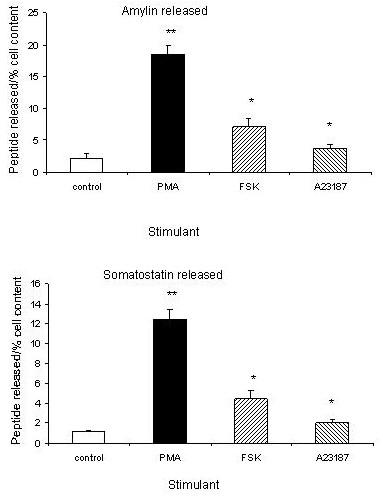
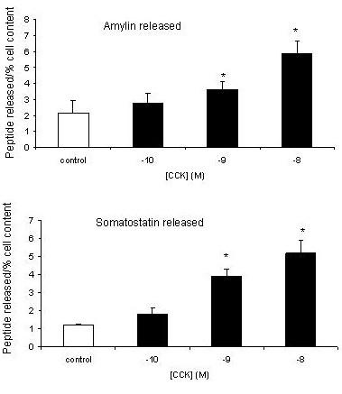
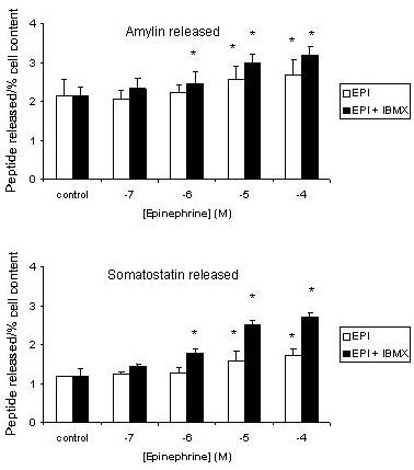
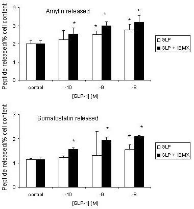
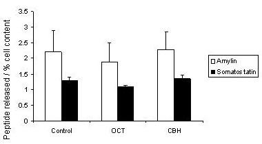
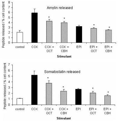
Results: Amylin release was significantly enhanced by activation of protein kinase C with phorbol-12-myristate-13-acetate, adenylate cyclase with forskolin and elevation of intracellular calcium with A23187. Cholecystokinin (CCK), epinephrine and glucagon-like peptide-1 (GLP-1) each stimulated amylin release in a dose-dependent manner. Maximal CCK-stimulated release was greater than either epinephrine or GLP-1, even when the effects of the latter two were enhanced by isobutylmethylxanthine. Stimulated amylin release was significantly inhibited by carbachol (by 51-59%) and octreotide (by 33-42%). Somatostatin release paralleled that of amylin.
Conclusions: The cultured D-cell model provides a means of studying amylin release. Amylin secretion is stimulated by receptor-dependent and -independent activation of Ca2+/protein kinase C and adenylate cyclase pathways. Inhibition involves activation of muscarinic receptors and auto-regulation by somatostatin.
Figures
References
-
- Westermark P, Wernstedt C, Wilander E, Sletten K. A novel peptide in the calcitonin gene related peptide family as an amyloid fibril protein in the endocrine pancreas. Biochem Biophys Res Commun. 1986;140:827–31. - PubMed
-
- Miyazato M, Nakazato M, Shiomi K, Aburaya J, Toshimori H, Kangawa K, Matsuo H, Matsukura S. Identification and characterization of islet amyloid polypeptide in mammalian gastrointestinal tract. Biochem Biophys Res Commun. 1991;181:293–300. - PubMed
-
- Lukinius A, Wilander E, Westermark GT, Engstrom U, Westermark P. Co-localization of islet amyloid polypeptide and insulin in the B cell secretory granules of the human pancreatic islets. Diabetologia. 1989;32:240–4. - PubMed
Publication types
MeSH terms
Substances
LinkOut - more resources
Full Text Sources
Miscellaneous
