. 2003 Nov;71(11):6243-55.
doi: 10.1128/IAI.71.11.6243-6255.2003.

Pathogenic Acanthamoeba spp secrete a mannose-induced cytolytic protein that correlates with the ability to cause disease

Affiliations

Pathogenic Acanthamoeba spp secrete a mannose-induced cytolytic protein that correlates with the ability to cause disease

Michael Hurt et al. Infect Immun. 2003 Nov.

Abstract

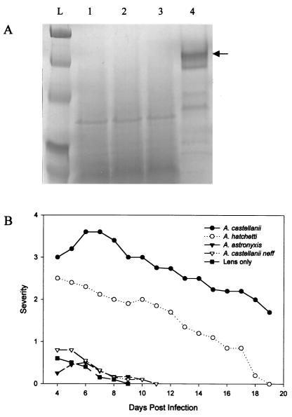
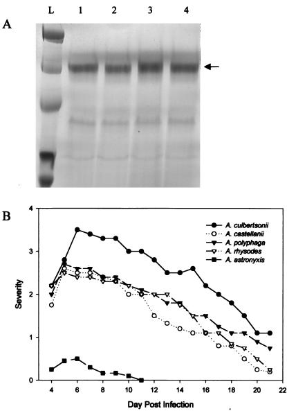
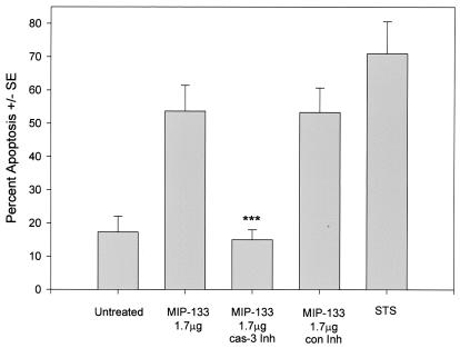
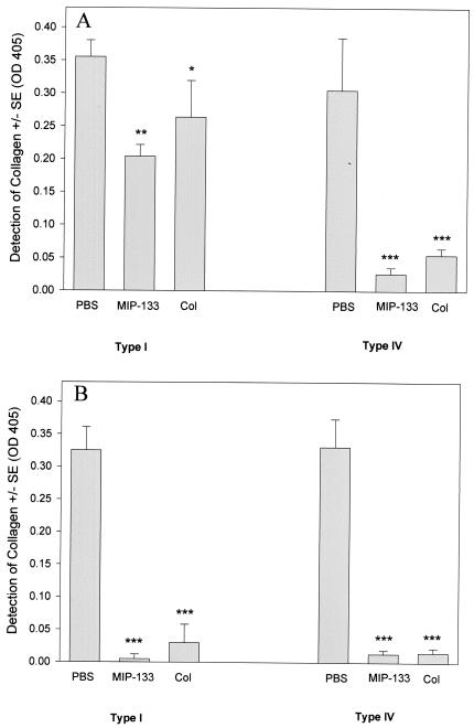
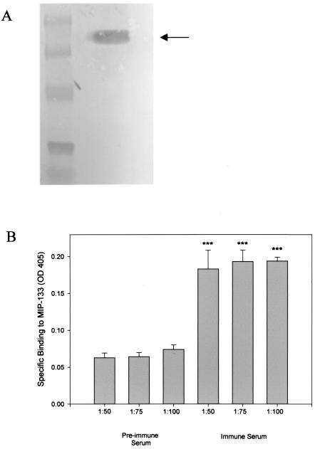
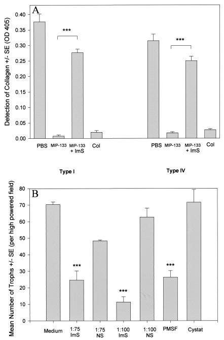
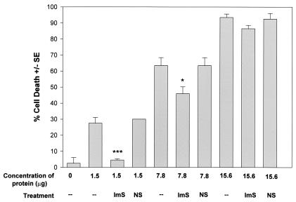
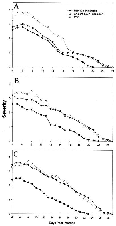
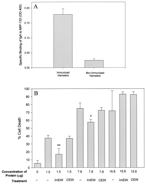
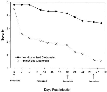
The pathogenesis of Acanthamoeba keratitis begins when Acanthamoeba trophozoites bind specifically to mannosylated glycoproteins upregulated on the surfaces of traumatized corneal epithelial cells. When Acanthamoeba castellanii trophozoites are grown in methyl-alpha-D-mannopyranoside, they are induced to secrete a novel 133-kDa protein that is cytolytic to corneal epithelial cells. Clinical isolates of Acanthamoeba spp., and not the soil isolates, were proficient at producing a mannose-induced protein (MIP-133) and generating disease in Chinese hamsters. The purified protein was efficient at killing corneal epithelial cells, the first mechanistic barrier, by inducing apoptosis in a caspase 3-dependent pathway. Subsequent steps in pathogenesis require the amoebae to penetrate and degrade collagen. Only the clinical isolates tested were efficient at migrating through a collagenous matrix in vitro, presumably by MIP-133 degradation of both human type I and human type IV collagen. A chicken anti-MIP-133 antiserum effectively bound to the protein and blocked collagenolytic activity, migration, and cytopathic effects (CPE) against corneal cells in vitro. Chinese hamsters orally immunized with MIP-133 displayed a >30% reduction in disease. Immunoglobulin A isolated from immunized animals bound MIP-133 and blocked CPE on corneal cells in vitro. Animals induced to generate severe chronic infections displayed significant reductions in disease symptoms upon oral immunization postinfection. These data suggest that MIP-133 production might be necessary to initiate corneal disease and that it may play an important role in the subsequent steps of the pathogenic cascade of Acanthamoeba keratitis. Furthermore, as antibodies produced both prior to and after infection reduced clinical symptoms of disease, the protein may represent an important immunotherapeutic target for Acanthamoeba keratitis.


Figures

FIG. 1.
FIG. 1.
Correlation between MIP-133 production and the ability of Acanthamoeba soil isolates to cause disease. (A) Acanthamoeba spp. were grown in PYG containing 100 mM methyl-α-d-mannopyranoside. Supernatants were collected at mid-log phase, filter sterilized, concentrated 10-fold, and analyzed by SDS-PAGE. Lanes: L, molecular weight ladder; 1, A. hatchetti; 2, A. astronyxis; 3, A. castellanii neff; 4, A. castellanii (clinical isolate). The arrow points at ∼133 kDa. (B) Ability of Acanthamoeba spp. to cause keratitis in Chinese hamsters. Animals were infected with Acanthamoeba-laden lenses as described in Materials and Methods. The results are representative of three separate experiments (eight hamsters in each group).
FIG. 2.
FIG. 2.
Correlation between MIP-133 production and the ability of clinical isolates of Acanthamoeba to cause disease. (A) Acanthamoeba spp. were grown in PYG containing 100 mM methyl-α-d-mannopyranoside. Supernatants were collected at mid-log phase, filter sterilized, concentrated 10-fold, and analyzed by SDS-PAGE. Lanes: L, molecular weight ladder; 1, A. culbertsoni; 2, A. polyphaga; 3, A. rhysodes; 4, A. castellanii. The arrow points at ∼133 kDa. (B) Ability of Acanthamoeba spp. to cause keratitis in Chinese hamsters. Animals were infected with Acanthamoeba-laden lenses as described in Materials and Methods. The results are representative of three separate experiments (eight hamsters in each group).
FIG. 3.
FIG. 3.
Effects of caspase 3 inhibition of MIP-133-mediated apoptosis. HCE cells were treated with 1.7 μg of MIP-133 either alone or with a 20 μM concentration of the caspase 3 inhibitor Z-DEVD-FMK (cas-3 Inh), or a 20 μM concentration of the caspase 3 control inhibitor Z-FA-FMK (con Inh). HCE cells were examined by Annexin V flow cytometry as described in Materials and Methods. Additional controls included untreated cells, and 3 μg of staurosporine (STS) per ml served as a positive control. Bars and error bars represent the means ± standard errors (SE) of results of triplicate experiments. ***, significantly different from the value for the untreated controls (P < 0.001).
FIG. 4.
FIG. 4.
Collagenolytic activity of the MIP-133 protein. Ten micrograms of human collagen types I and IV were incubated and dried onto 96-well plates. Wells were treated with either 15.6 μg of the MIP-133 protein, PBS, or 0.1 mg of collagenase (Col) for 24 (A) and 72 (B) h. Sample wells were then washed three times and incubated with mouse anti-collagen type IV IgG or mouse anti-collagen type I IgG as the primary antibody, followed by goat anti-mouse IgG-HRP, as described in Materials and Methods. Plates were developed and read at an OD at 405 nm. Bars and error bars represent the means ± SE of triplicate experiments. *, **, and ***, significantly different from values for PBS-treated controls (P < 0.05, P < 0.01, and P < 0.001, respectively).
FIG. 5.
FIG. 5.
Specific binding of the chicken anti-MIP-133 antiserum. (A) Western blotting. Crude supernatants taken from A. castellanii trophozoites grown in mannose were resolved by SDS-PAGE and transferred to PVDF membranes. Membranes were first incubated with chicken anti-MIP-133 antiserum, followed by rabbit anti-chicken IgG-alkaline phosphatase, as described in Materials and Methods. The membranes were then washed and developed with Nitro Blue Tetrazolium-BCIP stock solution. The arrow indicates the 133-kDa band. (B) ELISA. Ninety-six-well plates were coated with 50 μg of MIP-133 and allowed to dry. After drying, wells were blocked and incubated with a 1:50, 1:75, or 1:100 dilution of either the chicken anti-MIP-133 antiserum or preimmune chicken antiserum. Wells were then washed and incubated with goat anti-chicken IgY-HRP. ELISA plates were developed and read at an OD at 405 nm. Bars and error bars represent the means ± SE of results from triplicate experiments. ***, significantly different from values obtained with preimmune serum at the same dilutions (P < 0.001).
FIG. 6.
FIG. 6.
Anti-MIP-133 inhibition of MIP-133 collagenolytic activity and trophozoite migration. (A) ELISA. Ten-microgram samples of human collagen types I and IV were incubated and dried onto 96-well plates. Wells were treated with either 15.6 μg of the MIP-133 protein, 15.6 μg of the MIP-133 protein coincubated with a 1:75 dilution of chicken anti-MIP-133 antiserum (ImS), PBS, or 0.1 mg of collagenase (Col) for 72 h. Sample wells were then washed three times and incubated with mouse anti-collagen type IV IgG or mouse anti-collagen type I IgG as the primary antibody, followed by goat anti-mouse IgG-HRP, as described in Materials and Methods. Plates were then read at an OD at 405 nm. ***, significantly different from values from MIP-133 treatments (P < 0.001). (B) Migration assay. A. castellanii trophozoites (105) were placed in the upper chambers of 3.0-μm-pore-size transwells coated with Matrigel. Trophozoites were treated with 1:75 and 1:100 dilutions of either the chicken anti-MIP-133 antiserum (ImS) or the preimmune normal serum control (NS). Additional controls included 1.0 mM PMSF and 10 μM cystatin (Cystat). After 2 h, trophozoites in the bottom chamber were counted. Bars and error bars indicate means ± SE of results from 10 random high-powered fields (magnification, ×100). ***, significantly different from values obtained with the medium control or the normal serum control (P < 0.001).
FIG. 7.
FIG. 7.
Inhibition of MIP-133-mediated CPE against HCE cells. MIP-133 protein samples were adjusted to 1.5, 7.8, and 15.6 μg of protein in 25 μl of PBS before addition to HCE cells in 96-well microtiter plates for 18 h. Protein samples were either used alone, or coincubated with a 1:75 dilution of chicken anti-MIP-133 antiserum (ImS) or the normal serum control (NS). All final volumes were 200 μl. CPE were assessed spectrophotometrically. Each bar and error bar show the mean ± SE of triplicate counts. * and ***, significantly different from values for untreated controls (P < 0.05 and P < 0.001, respectively).
FIG. 8.
FIG. 8.
Effect of oral immunization with the MIP-133 protein on Acanthamoeba keratitis. Hamsters were orally immunized with either 100 (A), 200 (B), or 400 (C) μg of the MIP-133 protein once a week for 4 weeks prior to infection with A. castellanii-infected lenses as described in Materials and Methods. Lenses were removed 4 days postinfection, and corneas were evaluated for clinical severity at the times indicated. At all time points, the animals immunized with 400 μg of MIP-133 (C) displayed infections significantly different (P < 0.01) than those of the cholera toxin and PBS control groups. The results shown are representative of three separate experiments for each treatment (eight hamsters in each treatment group per experiment).
FIG. 9.
FIG. 9.
Inhibition of MIP-133-mediated CPE by mucosal anti-MIP-133 antibodies from immunized Chinese hamsters. (A) ELISA. Ninety-six-well plates were coated with 50 μg of MIP-133 and allowed to dry in carbonate buffer. After drying, wells were blocked and incubated with a 1:2 dilution of pooled enteric wash from immunized hamsters. Wells were then washed and incubated with a 1:2 dilution of rabbit anti-Chinese hamster IgA hyperimmune serum, washed, and further incubated with a 1:1,000 dilution of goat anti-rabbit IgG-HRP. ELISA plates were developed and read at an OD at 405 nm. (B) Inhibition of CPE. MIP-133 protein samples were adjusted to 1.5, 7.8, and 15.6 μg of protein in 25 μl of PBS before addition to HCE cells in 96-well microtiter plates for 18 h. Protein samples were either used alone or coincubated with a 1:50 dilution of pooled IgA enteric wash from immunized hamsters (ImEW) or of control enteric wash from PBS-immunized animals (CEW). All final volumes were 200 μl. CPE were assessed spectrophotometrically. Each bar and error bar show the mean ± SE of triplicate counts. * and **, significantly different from values for untreated controls (P < 0.05 and P < 0.01, respectively).
FIG. 10.
FIG. 10.
Effects of oral immunization in animals with chronic, progressive Acanthamoeba keratitis. Chinese hamsters were treated with clodronate-encapsulated liposomes administered via subconjunctival injection on days −8, −6, −4, and −2 prior to infection with A. castellanii-infected lenses. Hamsters were then either left untreated or orally immunized with 400 μg of the MIP-133 protein on days 5, 12, 19, and 26 postinfection. Lenses were removed 5 days postinfection, and corneas were evaluated for clinical severity at the times indicated. All time points of the immunized clodronate-treated animals displayed infections that were significantly different (P < 0.001) from those of the nonimmunized clodronate group after day 7 postinfection. The results shown are representative of two separate experiments for each treatment (eight hamsters in each group).

Similar articles

Cited by

References

    1. Alizadeh, H., S. Apte, M. S. El-Agha, L. Li, M. Hurt, K. Howard, H. D. Cavanagh, J. P. McCulley, and J. Y. Niederkorn. 2001. Tear IgA and serum IgG antibodies against Acanthamoeba in patients with Acanthamoeba keratitis. Cornea 20:622-627. - PubMed
    1. Alizadeh, H., J. Y. Niederkorn, and J. P. McCulley. 1996. Acanthamoeba keratitis, p. 1062-1071. In J. S. Pepose, G. N. Holland, and K. R. Wilhelmus (ed.), Ocular infection and immunity. Mosby, St. Louis, Mo.
    1. Auran, J. D., M. B. Starr, and F. A. Jacobiec. 1987. Acanthamoeba keratitis. Cornea 6:2-26. - PubMed
    1. Brown, T. J., R. T. M. Cursons, and E. A. Keys. 1982. Amoeba from antarctic soil and water. Appl. Environ. Microbiol. 44:491-493. - PMC - PubMed
    1. Centers for Disease Control. 1987. Acanthamoeba keratitis in soft-contact-lens wearers—United States. Morb. Mortal. Wkly. Rep. 36:397-398, 403-404. - PubMed

Publication types