. 2003 Nov;71(11):6344-53.
doi: 10.1128/IAI.71.11.6344-6353.2003.

Critical role of the complement system in group B streptococcus-induced tumor necrosis factor alpha release

Affiliations

Critical role of the complement system in group B streptococcus-induced tumor necrosis factor alpha release

Ofer Levy et al. Infect Immun. 2003 Nov.

Abstract

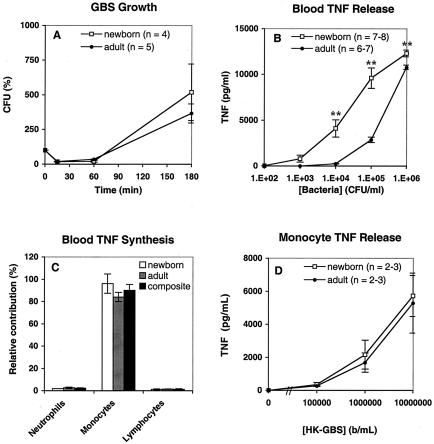
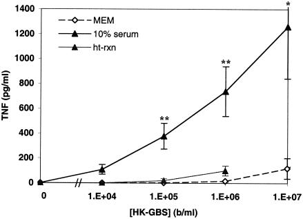
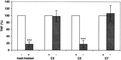
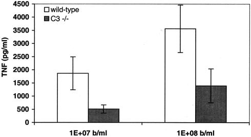
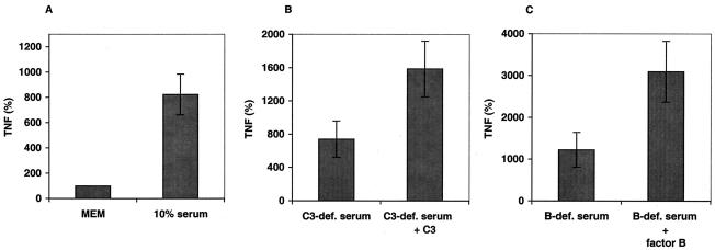
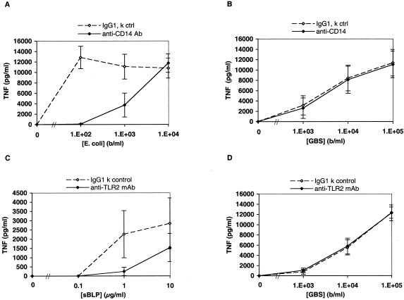
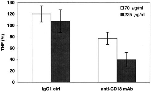
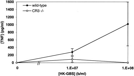
Group B Streptococcus (GBS) is a major cause of newborn sepsis and meningitis and induces systemic release of tumor necrosis factor alpha (TNF-alpha), believed to play a role in morbidity and mortality. While previous studies have shown that GBS can induce TNF-alpha release from monocytes and macrophages, little is known about the potential modulating effect of plasma or serum on GBS-induced TNF-alpha release, and there are conflicting reports as to the host receptors involved. In a human whole-blood assay system, GBS type III COH-1 potently induced substantial monocyte TNF-alpha release in adult peripheral blood and, due to a higher concentration of monocytes, 10-fold-greater TNF-alpha release in newborn cord blood. Remarkably, GBS-induced TNF-alpha release from human monocytes was enhanced approximately 1000-fold by heat-labile serum components. Experiments employing C2-, C3-, or C7-depleted serum demonstrated that C3 activation via the alternative pathway is crucial for potent GBS-induced TNF-alpha release. Accordingly, whole blood from C3-deficient mice demonstrated significantly reduced GBS-induced TNF-alpha release. Preincubation with human serum enhanced the TNF-alpha-inducing activity of GBS in a C3- and factor B-dependent manner, implying deposition of complement components via the alternative pathway. GBS-induced TNF-alpha release was inhibited by monoclonal antibodies directed against each of the components of CR3 and CR4: the common integrin beta subunit CD18 and the alpha subunits CD11b (of CR3) and CD11c (of CR4). Blood derived from CR3 (CD11b/CD18)-deficient mice demonstrated a markedly diminished TNF-alpha response to GBS. We conclude that the ability of plasma and serum to greatly amplify GBS-induced TNF-alpha release reflects the activity of the alternative complement pathway that deposits fragments on GBS and thereby enhances CR3- and CR4-mediated monocyte activation.


Figures

FIG. 1.
FIG. 1.
GBS induces TNF-α release from monocytes in newborn and adult blood. (A) Growth of GBS type III COH-1 added at 104 bacteria/ml to citrated adult blood (closed symbols) or newborn cord blood (open symbols) and incubated at 37°C. Bacterial growth was assessed by quantitative culture at the time indicated. (B) Extracellular medium was collected at 5 h, and TNF-α was measured by ELISA. TNF-α release is plotted relative to input inoculum (102 to 106 bacteria/ml). The total number of subjects studied (n) is indicated in each graph. Data points represent means ± standard errors of the means. P values were calculated for the comparison of TNF-α release in newborn and adult blood (**, P < 0.01). (C) Analysis of TNF-α-positive leukocytes derived from newborn blood (n = 2) and adult blood (n = 2) revealed that monocytes accounted for the greatest proportion of TNF-α synthesis. Differences between the relative contributions of composite monocytes and composite neutrophils or lymphocytes were significant (P < 0.001). (D) HK-GBS was added to neonatal or adult monocytes suspended to 105 bacteria/ml in 10% autologous serum. After a 5-h incubation at 37°C, the supernatants were collected for TNF-α measurement.
FIG. 2.
FIG. 2.
Heat-labile serum components potentiate GBS-induced TNF-α release from human monocytes in culture. PBMC-derived monocytes were cultured in MEM, 10% fresh autologous serum (in MEM), or 10% heat-treated autologous serum, after which HK-GBS was added at the indicated concentrations. TNF-α release was measured by ELISA. Similar results were obtained with newborn cord blood- and adult peripheral blood-derived monocytes, and data were pooled. Data represent the means ± standard errors of the means of 2 to 22 independent determinations. Differences between serum and MEM (*, P < 0.05; **, P < 0.01) as well as differences between serum and heat-treated serum (P < 0.01) were significant.
FIG. 3.
FIG. 3.
Heat treatment or depletion of C3, but not of C2 or C7, reduces the ability of human serum to potentiate GBS-induced TNF-α release from human monocytes. HK-GBS (106 bacteria/ml) was added to cultured monocytes in the presence of 10% heat-treated autologous serum or 10% C2-, C3-, or C7-depleted heterologous pooled serum. After a 5-h incubation, the extracellular medium was harvested for TNF-α measurement. The data are presented as pairwise comparisons to the “normal” or repleted condition that is normalized to 100% TNF-α release. Similar results were obtained with newborn and adult monocytes, and the data were pooled (n = 3 to 9). Differences between normal serum and heat-treated serum and between C3-depleted and C3-repleted serum were significant (***, P < 0.001).
FIG. 4.
FIG. 4.
Whole blood from mice deficient in complement component C3 exhibits reduced GBS-induced TNF-α release. Peripheral blood of healthy sex- and age-matched control C57BL/6 mice or C3-deficient mice was incubated with the indicated concentrations of HK-GBS for 5 h before collection of the extracellular fluid for measurement of murine TNF-α by ELISA. Background TNF-α release in control (i.e., no GBS) samples was subtracted to obtain GBS-induced TNF-α release. Data represent the means ± standard errors of the means (n = 6). Differences between wild-type and C3−/− mice were significant (P < 0.05).
FIG. 5.
FIG. 5.
TNF-α-inducing activity of GBS is markedly enhanced by preexposure to serum in a C3- and factor B-dependent manner. HK-GBS was preincubated for 1 h at 37°C in MEM alone (MEM) or MEM supplemented with 10% serum (A), 10% C3-depleted serum with or without repletion of C3 (B), or 10% factor B-depleted serum with or without repletion of factor B (C). Purified complement components were added to final concentrations found in 10% serum. After preincubation, bacteria were washed three times with PBS and added to cultured monocytes at a concentration of 106 or 107 bacteria per ml. After a 5-h incubation, the extracellular medium was harvested for TNF-α measurement. Data are normalized as percentages of the TNF-α-inducing activity of HK-GBS incubated in MEM alone (defined as 100%) and represent the means ± standard errors of the means. Similar results were obtained with newborn and adult monocytes, and the data were pooled (n = 6 to 15). Differences between MEM and 10% serum (P < 0.01), C3-depleted serum and C3-repleted serum (P < 0.05), and factor B-depleted and factor B-repleted serum (P < 0.05) were significant.
FIG. 6.
FIG. 6.
Neutralizing MAbs to CD14 and TLR2 inhibit ligand-induced TNF-α release in whole blood but do not inhibit GBS-induced TNF-α release. The figure shows the effect of preincubation of newborn or adult whole blood (pooled data) with an isotype control antibody or a neutralizing MAb to CD14 on TNF-α release induced by live E. coli K1/r (A) or GBS type III COH-1 (B). Differences in the effects of the CD14 MAb between E. coli and GBS were significant (n = 4 to 8; P < 0.01). To evaluate TLR2, newborn or adult blood was preincubated with 150 μg of an isotype control MAb or the neutralizing TLR2 MAb 2392 per ml, after which the known TLR2 agonist sBLP (C) or L-GBS (D) was added. Differences in the effects of the TLR2 MAb between sBLP and GBS were significant (n = 4, P < 0.001).
FIG. 7.
FIG. 7.
A neutralizing CD18 MAb inhibits GBS-induced TNF-α release. Newborn or adult blood was preincubated for 1 h with either a control murine IgG1 MAb to an unrelated antigen (antitrinitrophenol) or a neutralizing murine IgG1 MAb to CD18 before addition of L-GBS type III COH-1 (104 bacteria/ml). TNF-α release was measured at 5 h by ELISA and is expressed as a percentage of control. Data represent the means ± standard errors of the means (n = 2 to 4). Differences between control samples and those containing 225 μg of CD18 MAb/ml were significant (P < 0.05).
FIG. 8.
FIG. 8.
Blood derived from mice deficient in the complement receptor CR3 demonstrates markedly reduced GBS-induced TNF-α release. Blood from CR3-deficient or age- and sex-matched control C57/129 mice was incubated with HK-GBS at 107 or 108 bacteria/ml before measurement of TNF-α by ELISA. Data represent the means ± standard errors of the means (n = 4 to 6). Differences between CR3-deficient and control mice were significant (P < 0.05, Fisher's exact test).

Similar articles

Cited by

References

    1. Aderem, A., and R. J. Ulevitch. 2000. Toll-like receptors in the induction of the innate immune response. Nature 406:782-787. - PubMed
    1. Albanyan, E. A., and M. S. Edwards. 2000. Lectin site interaction with capsular polysaccharide mediates nonimmune phagocytosis of type III group B streptococci. Infect. Immun. 68:5794-5802. - PMC - PubMed
    1. Albanyan, E. A., J. G. Vallejo, C. W. Smith, and M. S. Edwards. 2000. Nonopsonic binding of type III group B streptococci to human neutrophils induces interleukin-8 release mediated by the p38 mitogen-activated protein kinase pathway. Infect. Immun. 68:2053-2060. - PMC - PubMed
    1. Aliprantis, A. O., R. B. Yang, M. R. Mark, S. Suggett, B. Devaux, J. D. Radolf, G. R. Klimpel, P. Godowski, and A. Zychlinsky. 1999. Cell activation and apoptosis by bacterial lipoproteins through toll-like receptor-2. Science 285:736-739. - PubMed
    1. Baker, C. J., M. S. Edwards, B. J. Webb, and D. L. Kasper. 1982. Antibody-independent classical pathway-mediated opsonophagocytosis of type Ia, group B streptococcus. J. Clin. Investig. 69:394-404. - PMC - PubMed

Publication types