

Pertussis toxin plays an early role in respiratory tract colonization by Bordetella pertussis
- PMID: 14573656
- PMCID: PMC219603
- DOI: 10.1128/IAI.71.11.6358-6366.2003
Pertussis toxin plays an early role in respiratory tract colonization by Bordetella pertussis
Abstract
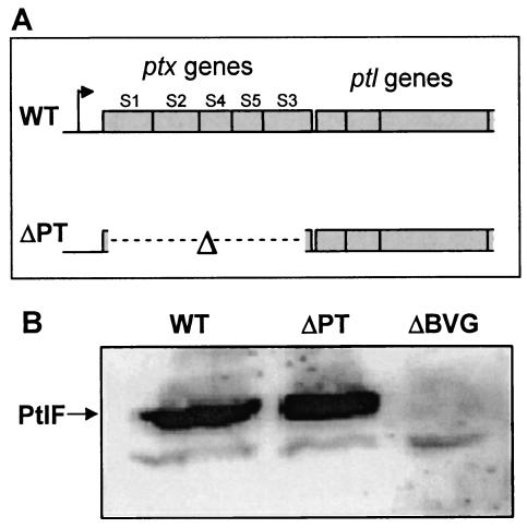
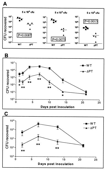
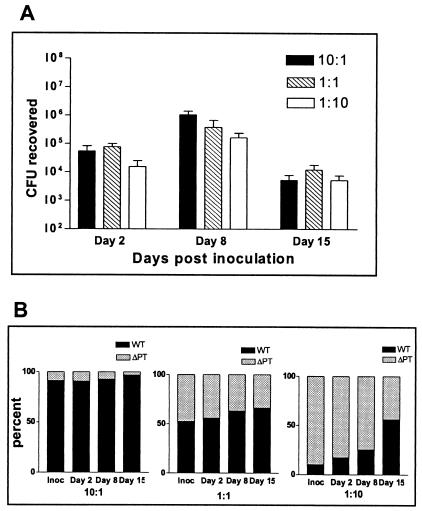
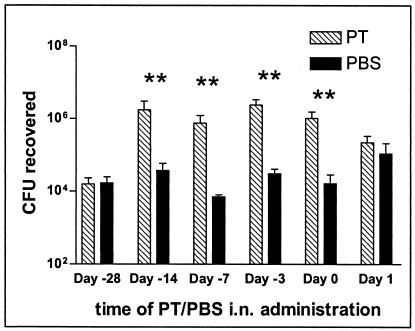
In this study, we sought to determine whether pertussis toxin (PT), an exotoxin virulence factor produced exclusively by Bordetella pertussis, is important for colonization of the respiratory tract by this pathogen by using a mouse intranasal infection model. By comparing a wild-type Tohama I strain to a mutant strain with an in-frame deletion of the ptx genes encoding PT (deltaPT), we found that the lack of PT confers a significant peak (day 7) colonization defect (1 to 2 log(10) units) over a range of bacterial inoculum doses and that this defect was apparent within 1 to 2 days postinoculation. In mixed-strain infection experiments, the deltaPT strain showed no competitive disadvantage versus the wild-type strain and colonized at higher levels than in the single-strain infection experiments. To test the hypothesis that soluble PT produced by the wild-type strain in mixed infections enhanced respiratory tract colonization by deltaPT, we coadministered purified PT with the deltaPT inoculum and found that colonization was increased to wild-type levels. This effect was not observed when PT was coadministered via a systemic route. Intranasal administration of purified PT up to 14 days prior to inoculation with deltaPT significantly increased bacterial colonization, but PT administration 1 day after bacterial inoculation did not enhance colonization versus a phosphate-buffered saline control. Analysis of bronchoalveolar lavage fluid samples from mice infected with either wild-type or deltaPT strains at early times after infection revealed that neutrophil influx to the lungs 48 h postinfection was significantly greater in response to deltaPT infection, implicating neutrophil chemotaxis as a possible target of PT activity promoting B. pertussis colonization of the respiratory tract.
Figures
Similar articles
-
Suppression of serum antibody responses by pertussis toxin after respiratory tract colonization by Bordetella pertussis and identification of an immunodominant lipoprotein.Infect Immun. 2004 Jun;72(6):3350-8. doi: 10.1128/IAI.72.6.3350-3358.2004. Infect Immun. 2004. PMID: 15155640 Free PMC article.
-
Pertussis toxin and adenylate cyclase toxin provide a one-two punch for establishment of Bordetella pertussis infection of the respiratory tract.Infect Immun. 2005 May;73(5):2698-703. doi: 10.1128/IAI.73.5.2698-2703.2005. Infect Immun. 2005. PMID: 15845471 Free PMC article.
-
Pertussis toxin exacerbates and prolongs airway inflammatory responses during Bordetella pertussis infection.Infect Immun. 2012 Dec;80(12):4317-32. doi: 10.1128/IAI.00808-12. Epub 2012 Oct 1. Infect Immun. 2012. PMID: 23027529 Free PMC article.
-
The concept of pertussis as a toxin-mediated disease.Pediatr Infect Dis. 1984 Sep-Oct;3(5):467-86. doi: 10.1097/00006454-198409000-00019. Pediatr Infect Dis. 1984. PMID: 6093069 Review.
-
Contribution of pertussis toxin to the pathogenesis of pertussis disease.Pathog Dis. 2015 Nov;73(8):ftv073. doi: 10.1093/femspd/ftv073. Epub 2015 Sep 21. Pathog Dis. 2015. PMID: 26394801 Free PMC article. Review.
Cited by
-
Genetically detoxified pertussis toxin displays near identical structure to its wild-type and exhibits robust immunogenicity.Commun Biol. 2020 Aug 5;3(1):427. doi: 10.1038/s42003-020-01153-3. Commun Biol. 2020. PMID: 32759959 Free PMC article.
-
Intracellular disassembly and activity of pertussis toxin require interaction with ATP.Pathog Dis. 2016 Aug;74(6):ftw065. doi: 10.1093/femspd/ftw065. Epub 2016 Jun 30. Pathog Dis. 2016. PMID: 27369899 Free PMC article.
-
Bordetella bronchiseptica and Bordetella pertussis: Similarities and Differences in Infection, Immuno-Modulation, and Vaccine Considerations.Clin Microbiol Rev. 2023 Sep 21;36(3):e0016422. doi: 10.1128/cmr.00164-22. Epub 2023 Jun 12. Clin Microbiol Rev. 2023. PMID: 37306571 Free PMC article. Review.
-
Pertussis toxin and adenylate cyclase toxin: key virulence factors of Bordetella pertussis and cell biology tools.Future Microbiol. 2010 Mar;5(3):455-69. doi: 10.2217/fmb.09.133. Future Microbiol. 2010. PMID: 20210554 Free PMC article. Review.
-
Role of neutrophils in response to Bordetella pertussis infection in mice.Infect Immun. 2009 Mar;77(3):1182-8. doi: 10.1128/IAI.01150-08. Epub 2008 Dec 22. Infect Immun. 2009. PMID: 19103765 Free PMC article.
References
-
- Brennan, M. J., J. L. David, J. G. Kenimer, and C. R. Manclark. 1988. Lectin-like binding of pertussis toxin to a 165 kilodalton Chinese hamster ovary cell glycoprotein. J. Biol. Chem. 263:4895-4899. - PubMed
-
- Carbonetti, N. H., T. J. Irish, C. H. Chen, C. B. O'Connell, G. A. Hadley, U. McNamara, R. G. Tuskan, and G. K. Lewis. 1999. Intracellular delivery of a cytolytic T-lymphocyte epitope peptide by pertussis toxin to major histocompatibility complex class I without involvement of the cytosolic class I antigen processing pathway. Infect. Immun. 67:602-607. - PMC - PubMed
Publication types
MeSH terms
Substances
Grants and funding
LinkOut - more resources
Full Text Sources
Other Literature Sources
Molecular Biology Databases
